About Myocardial Infarction
Myocardial infarction
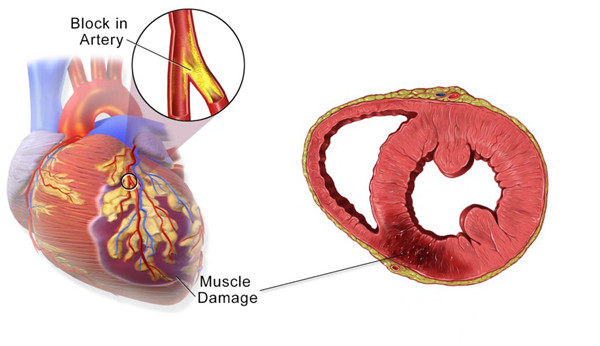
Myocardial infarction (MI) is myocardial ischemic necrosis, coronary artery occlusion, and interruption of blood flow, which causes local necrosis for part of the myocardium due to severe persistent ischemia. With the heart load increased, the blood flow of the coronary artery can not increase accordingly, and the myocardium is in a state of ischemia, resulting in the onset of myocardial infarction. 60%-89% of patients suffer from high blood pressure during the period of the disease, and nearly half of the patients have angina pectoris, and more men than women suffer with myocardial infarction due to sudden abnormal excitement or stimulation. Myocardial infarction is an emergency signal from the heart, indicating that the heart is anoxic and starts to have cardiac necrosis, which will cause a variety of symptoms. A myocardial infarction may cause heart failure, an irregular heartbeat, cardiac arrest or cardiogenic shock.

Figure 1: Myocardial Infarction (Sourcing:http://www.miracormedical.com/picso-therapy/picso-in-ami/clinical-background/)
Let's take acute myocardial infarction (AMI) as an example. At least 80% to 90% of people with MI have early signs, such as chest tightness, dull pain, or palpitations. However, the early symptoms only last a short time. They are often ignored by people because of the alleviation in minutes or dozens of minutes. The 90 minutes of early onset of acute myocardial infarction is the golden hour of treatment. Thus try your best to go to the hospital once you feel chest tightness and chest pain. The effect of treatment is the best in 3 hours after myocardial infarction. The longer the delay, the more serious the injury of the myocardium, and the greater the difficulty of treatment.
In recent years, the tragedy of sudden acute myocardial infarction leading to death is often reported. Among these people, many are young and middle-aged. The lack of exercise leads to significant thickening of blood vessel walls, and long-term smoking leads to abnormal blood lipids. Eating and sleeping irregularities, overwork, and overtime staying up a late lead to decreased body resistance. All these factors are easy to cause the onset of this disease. Most middle-aged and young people with acute myocardial infarction have different levels of basic diseases.
For myocardial infarction, in addition to routine blood tests and electrocardiogram tests, clinically assisted serological tests such as creatine kinase isozyme (CKMB protein), lactate dehydrogenase, myoglobin( myo antibody), troponin, N-terminal brain sodium peptide precursor(N terminal proBNP), and C-reactive protein (CRP) are often used in combination. Measuring cardiac biomarkers can be an assistant to diagnose cardiac diseases. Among the many biomarkers, troponin is considered to be a superior and preferred marker for measuring myocardial injury due to its sensitivity and specificity in tests compared to other tests. Troponin levels elevate within 2-3 hours after myocyte injury and peak within 1-2 days. Troponins can also calculate infarct size.
SEKBIO provide a series of antigens of Myocardial infarction including cTnl, Myo, CKMB, H-FABP, D-Dimer, NT-proBNP, ST-2.
Clinical correlation of flagship material pairs comparable to reagent values of internationally top brands.
Great batch-to-batch consistency.
Cardiac troponin I (cTnI)
| Catalog No. | Product Name | Clone | Epitope |
|---|---|---|---|
| ZLA1011M | Mouse anti-human cTnI, mAb | 1G4M | 41-49 |
| ZLA1011H | Recombinant anti- human cTnI, mAb | 1G4H | 41-49 |
| ZLA1012M | Mouse anti-human cTnI, mAb | 1B10M | 24-40 |
| ZLA1012H | Recombinant anti- human cTnI, mAb | 1B10H | 24-40 |
| ZLA1016M | Mouse anti-human cTnI, mAb | 5F3M | Complex |
| ZLA1017M | Mouse anti-human cTnI, mAb | 3G7M | 86-90 |
| ZLA1017H | Recombinant anti- human cTnI, mAb | 3G7H | 86-90 |
| ZLA1018M | Mouse anti-human cTnI, mAb | 10F9M | 169-178 |
| ZLA10110M | Mouse anti-human cTnI, mAb | 7B1M | Troponin C |
| ZLA10111M | Mouse anti-human cTnI, mAb | 4B6M | 83-93 |
| ZLP1011 | Recombinant human cardiac troponin IC complex I (cTnI-C-I) | Antigen | |
| ZLP1012 | Recombinant human cardiac troponin IC complex II (cTnI-C-II) | Antigen |
Myoglobin (Myo)
| Catalog No. | Product Name | Clone |
|---|---|---|
| ZLA1211M | Mouse anti-human Myo, mAb | 3H7M |
| ZLA1212M | Mouse anti-human Myo, mAb | 4D2M |
| ZLA1212H | Humanized anti-Myo, mAb | 4D2H |
Heart-type Fatty Acid Binding Protein (H-FABP)
H-FABP(heart-type fatty acid binding protein) is a low-molecular-weight cytosolic protein that is involved in the intracellular uptake and buffering of fatty acids in the myocardium. It is expressed primarily in the heart, but it is not 100% cardio specific since it is expressed to a lesser extent in skeletal muscle, brain, and kidneys. During myocardial injury, the H-FABP level in serum is elevated rapidly, making it an ideal marker for myocardial infarction, and it is a useful prognostic marker in patients with proven acute coronary syndrome. However, in renal failure and skeletal muscle disease, it has limited diagnostic value for AMI.
| Catalog No. | Product Name | Clone |
|---|---|---|
| ZLA1041M | Mouse anti-human FABP mAb | 9C5M |
| ZLA1042M | Mouse anti-human FABP mAb | 5D9M |
| ZLA1042H | Humanized anti-human FABP mAb | 5D9H |
| ZLP1041 | Recombinant Antigen (H-FABP) | Antigen |
D-Dimer
D-dimer (or D dimer) is a fibrin degradation product (or FDP), a small protein fragment present in the blood after a blood clot is degraded by fibrinolysis.
In order to eliminate blood clots from the body before they become problematic, a blot clot will go through Fibrinolysis; the process of degrading blood clot fibrin. A key product of fibrin degradation is the small protein fragment D-Dimer. Because of this link, D-Dimer is commonly used in the clinic as a biomarker of fibrin deposition and stabilization. Most often, the concentration of blood based D-Dimer is used to diagnose deep venous thrombosis (DVT), pulmonary embolism (PE) or disseminated intravascular coagulation (DIC) due to its strong association with blot clot formation.
| Catalog No. | Product Name | Clone |
|---|---|---|
| ZLA1051M | Mouse anti-human D-Dimer mAb | 2B3M |
| ZLA1052M | Mouse anti-human D-Dimer mAb | 4C1M |
| ZLA1052H | Humanized anti-D-Dimer mAb | 4C1H |
| ZLA1053M | Mouse anti-human D-Dimer mAb | 2A10M |
| ZLA1054M | Mouse anti-human D-Dimer mAb | 2G5M |
| ZLA1054H | Humanized anti-D-Dimer mAb | 2G5H |
| ZLA1055M | Mouse anti-human D-Dimer mAb | 2D6M |
N-terminal pro B type Natriuretic Peptide (NT-proBNP)
proBNP is secreted by myocardial cells in response to increased volume and pressure. This precursor molecule is cleaved to form the active BNP and the inactive N-terminal fragment of pro-BNP (NT-proBNP). Comparisons of BNP and NT-proBNP have shown that both molecules are effective in diagnosing left ventricular dysfunction in the acute care/emergency setting.
| Catalog No. | Product Name | Clone | Epitope |
|---|---|---|---|
| ZLA1021M | Mouse anti-human NT-proBNP mAb | 5D12M | 31-39 |
| ZLA1022M | Mouse anti-human NT-proBNP mAb | 5F1M | 13-24 |
| ZLA1023M | Mouse anti-human NT-proBNP mAb | 1H6M | 67-76 |
| ZLA1025M | Mouse anti-human NT-proBNP mAb | 4C7M | 27-31 |
| ZLA1026S | Sheep anti-human NT-proBNP mAb | 1B9S | 42-46 |
| ZLA1027M | Mouse anti-human NT-proBNP mAb | 5B12M | 39-48 |
| ZLA1027H | Humanized anti-NT-proBNP mAb | 5B12H | 39-48 |
| ZLP1023 | Recombinant Antigen (NT-proBNP ) |


