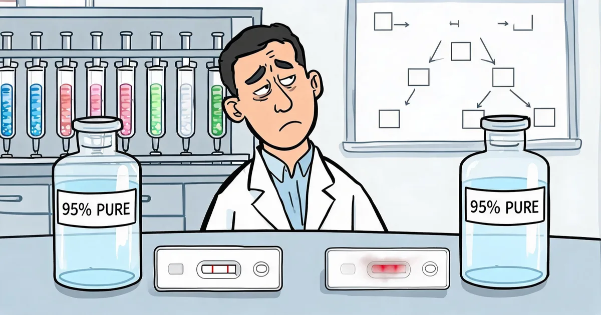
You order two lots of antibody against the same target. Both certificates of analysis read "≥95% purity, SDS-PAGE." One gives you a clean lateral flow strip. The other gives you a smeared test line, background across the control window, and a gold conjugate that flocculates overnight.
The purity number was accurate. SDS-PAGE separates proteins by molecular weight after denaturing them, so it tells you what is in the vial but not what state it is in. The remaining 5% could be harmless albumin. It could also be aggregates that wreck colloidal gold conjugation, leached Protein A that binds your sample matrix, or fragments that quietly dilute your effective titer.
Antibody purification is the set of separation steps that isolates immunoglobulin from serum, ascites, hybridoma supernatant or recombinant cell culture. Nearly every antibody reagent on the market passes through some combination of six methods, and which ones were used shows up later on your strip, your plate and your bridging studies.
Protein A, G and L Affinity Capture
Protein A affinity chromatography uses a cell-wall protein from Staphylococcus aureus that binds the Fc region of IgG at the CH2–CH3 interface. Immobilized on a resin, it captures IgG directly from almost any crude feed in one step. Elution is triggered by dropping the pH to about 2.5–3.5, which breaks the Fc–ligand interaction.
A single Protein A step typically delivers greater than 90% purity. Binding capacity is less predictable than resin brochures suggest. In a comparison of 15 commercial Protein A media, dynamic binding capacity depended strongly on residence time, and agarose-based sorbents only reached their higher capacity at residence times longer than 3 minutes (Hahn et al., 2003). Ask for capacity data at the flow rate the supplier actually runs.
Isotype compatibility decides the resin
The compatibility table matters more than any resin specification:
- Mouse IgG1, the most common hybridoma subclass, binds Protein A only weakly. Protein G is the safer capture choice.
- Mouse IgG2a and IgG2b bind both Protein A and Protein G well.
- Human IgG3, most rat IgG and chicken IgY do not bind Protein A.
- Fab and scFv fragments have no Fc region. Protein L, which binds kappa light chains, is the usual capture route.
Elution and ligand leaching
Low-pH elution is the acknowledged aggregation risk of Protein A capture (Vázquez-Rey & Lang, 2011). Raising the elution pH is gentler, but recovery falls. In one published comparison, 0.1 M citrate at pH 3.8 recovered less than 50% of bound monoclonal antibody, and recovery dropped further as pH rose to 4.3. At the same pH, 0.5 M and 2 M arginine eluents recovered much more, and the eluted antibody was mostly monomeric with either eluent (Arakawa et al., 2004). Fast neutralization after elution is standard process control.
The ligand itself also leaks. Trace Protein A in the final material can cause non-specific binding downstream, especially in formats that contain IgG from other species. Residual Protein A is usually measured by ELISA, and an antibody such as our chicken anti-Protein A antibody (IgY) is useful here because chicken IgY does not bind Protein A itself.
Three questions that predict background better than purity
Ask your supplier how they elute from Protein A, how quickly they neutralize, and what their residual Protein A specification is. The answers tell you more about background risk on your strip than the SDS-PAGE figure on the CoA.
Ion Exchange: Clearing Impurities and Charge Variants
Ion exchange chromatography (IEX) separates proteins by net charge at a given pH. The antibody and its impurities are matched against a positively charged anion exchanger (AEX) or a negatively charged cation exchanger (CEX), and bound proteins elute as salt concentration or pH changes.
Two modes dominate:
- AEX in flow-through mode. The antibody passes through while host-cell proteins, DNA, viruses and endotoxin bind. This is the standard impurity-clearance step after Protein A in recombinant processes.
- CEX in bind-elute mode. The antibody is retained and eluted with high resolution, removing aggregates, charge variants and leached Protein A.
For IVD developers the point is subtle. Charge heterogeneity is a hidden cause of lot-to-lot drift in assay performance, and a CEX polish narrows the charge-variant distribution of the final product. If two lots of the "same" antibody behave differently, ask whether the supplier runs a charge-homogenizing polish, and ask for the lot-to-lot titer CV. Our article on antibody batch consistency in IVD manufacturing covers which release parameters track kit performance.
HIC and SEC: Where Aggregates Get Removed
Hydrophobic interaction chromatography (HIC)
HIC separates by surface hydrophobicity. The antibody binds at high salt, commonly ammonium sulfate, and elutes as the salt concentration falls. Aggregates expose more hydrophobic surface than monomers, so they bind harder and elute later. That makes HIC a strong aggregate-removal step, and because it selects on a different property than ion exchange, the two pair well in a polishing train.
Size exclusion chromatography (SEC)
SEC, also called gel filtration, separates molecules by hydrodynamic radius as they pass through porous beads. Aggregates elute first; fragments and salts come out last. It is the gentlest method available: no binding chemistry, no pH extremes, no salt shock. It is also the slowest and lowest in capacity, so it is almost never used for capture.
SEC has three practical roles:
- Final polishing to remove aggregates and fragments.
- Buffer exchange into a conjugation-compatible formulation.
- Analytical SEC-HPLC, the QC method used to report monomer percentage.
For colloidal gold and latex conjugation, aggregate content matters as much as purity. Aggregates nucleate non-specific particle binding and cause flow defects. A monomer content of ≥95% by SEC-HPLC is a common conjugation-grade specification. If a supplier reports neither an HIC nor an SEC step and gives no SEC-HPLC data, treat aggregate content as unknown. Our troubleshooting guide to colloidal gold conjugation mistakes in rapid tests covers what else can go wrong at that step.
Precipitation for Serum Polyclonals
Precipitation purifies antibodies without a column, by fractionating on solubility. Caprylic (octanoic) acid at mildly acidic pH precipitates albumin and most non-immunoglobulin serum proteins. Ammonium sulfate then salts out and concentrates the IgG.
The two-step caprylic acid and ammonium sulfate protocol has been a standard route to serum IgG since it was described for serum and ascites in 1987 (McKinney & Parkinson, 1987). It is cheap, needs no chromatography equipment, scales to liters of serum and works across most mammalian sera. It remains a common first step for bulk polyclonal IgG, including many lateral flow coating antibodies.
Its limits are equally clear. Purity tops out at roughly 80–90%, and serum biology varies between bleeds and between animals. Precipitation alone is fine for coating antibodies. For conjugation-grade material, pair it with an affinity or ion exchange polish, and ask how the supplier manages serum-source variability between lots.
Magnetic Beads and Membranes at Small Scale
Magnetic bead purification immobilizes Protein A, G or L, or recombinant single-domain (VHH) affinity ligands, on superparamagnetic particles. Binding, washing and elution happen in a tube with a magnet. There are no columns and no centrifugation, and the format automates easily.
Beads dominate microgram-to-milligram purification and high-throughput clone screening. The ligand chemistry is changing in a direction IVD manufacturers should watch. Recombinant VHH ligands (12–15 kDa fragments produced in yeast) are animal-origin-free, and raw material origin is an increasingly common question in regulatory files.
Membrane and monolith formats use convective flow through pores instead of diffusion into beads, which allows high flow rates at low pressure.
Choosing a Purification Train by Source and Application
No single method produces a finished reagent. Real processes are trains: a capture step chosen by antibody source, followed by polish steps chosen by application. Start with the source.
| Antibody source | Typical workflow | Why |
|---|---|---|
| Serum or ascites (polyclonal) | Caprylic acid + ammonium sulfate → Protein A/G polish → IEX or SEC | High volume at low cost; polish only where conjugation-grade material is needed |
| Hybridoma supernatant | Protein G (IgG1) or Protein A (IgG2a/2b) → IEX polish | Capture resin must match the subclass |
| Recombinant CHO or HEK293 | Protein A → AEX flow-through → CEX or HIC polish | The platform process; lowest host-cell protein and DNA in two to three steps |
| E. coli periplasm (Fab, scFv) | Protein L or IMAC (His-tag) → SEC | No Fc region; endotoxin control becomes mandatory |
| IgM (any source) | Euglobulin precipitation → IEX → SEC; Protein A/G not suitable | Pentameric IgM is large and fragile, and binds poorly to IgG ligands |
IgM is a special case with its own failure modes. If you work with IgM capture antibodies, see our IgM purification services and the companion article on IgM purification mistakes.
Then filter by application:
- Lateral flow conjugation (colloidal gold, latex): aggregate removal and buffer composition decide success. Specify SEC-HPLC monomer ≥95% and a preservative-free, low-ionic-strength buffer.
- ELISA coating: purity demands are moderate. Titer and specificity carry the assay.
- Enzyme conjugation: sodium azide inhibits HRP, so the antibody must be azide-free before labeling. Our HRP conjugation mistakes article covers the rest of that checklist.
- IHC and immunofluorescence: consider Fab fragments to reduce Fc-mediated background.
- Anything that touches live cells: endotoxin specifications apply.
Scale is the last filter: beads and spin columns for micrograms to milligrams, packed columns for milligrams to grams, process chromatography beyond that.
What a Purity Number Leaves Out
"95% pure" is a claim about composition. Quality is a claim about fitness for your application. Three questions close the gap.
What is the other 5%? Albumin is largely harmless for coating. Aggregates ruin gold conjugation. Leached Protein A creates non-specific binding. Fragments dilute titer. The identity of the impurity matters more than its size.
Which method measured the purity? Denaturing SDS-PAGE cannot see aggregates. Native SEC-HPLC can, and it is the number that predicts conjugation behavior.
What does lot-to-lot consistency look like? For an IVD manufacturer, this is the real question. Ask for titer CV across recent lots, the SEC-HPLC monomer trend and, for polyclonals, how immunization and bleed pooling are standardized. Sequence-defined recombinant antibodies from a CHO stable cell line remove most of that biological variability at the source.
A seven-point antibody supplier checklist
(1) SEC-HPLC monomer % and method; (2) purity method, reducing or non-reducing SDS-PAGE or SEC-HPLC; (3) residual Protein A/G; (4) host-cell protein and residual DNA for recombinant material; (5) endotoxin level; (6) buffer composition and preservatives, since azide blocks HRP conjugation and preservatives interfere with particle coupling; (7) lot-to-lot titer CV.
Five Common Failure Modes and Their Upstream Causes
| Symptom at your bench | Upstream cause | Where to look |
|---|---|---|
| Gold conjugate flocculates; high background on strip | Aggregates; preservative in formulation buffer | SEC-HPLC monomer ≥95%; preservative-free, low-salt buffer |
| Antibody lost activity after purification | Harsh low-pH elution during Protein A capture | Milder elution (pH 3.5–4), arginine eluent, immediate neutralization |
| Non-specific binding in ELISA | Leached Protein A; polyclonal cross-reactivity; heterophilic interference in samples | Residual ligand specification; antigen-affinity polish of pAb; heterophilic blocking reagent |
| Weak test line despite good titer on paper | Fragments or charge variants; capture resin mismatched to subclass | SEC or CEX polish; verify resin–subclass compatibility |
| Lot fails bridging study | Charge heterogeneity; serum-source drift between bleeds | CEX polish; standardized immunization and pooling; reserve-lot bridging |
If a lot is drifting, start the diagnosis upstream. A quick functional check through express antibody validation can tell you whether the problem sits in the antibody or in your assay conditions.
Frequently Asked Questions
Protein A or Protein G for a mouse hybridoma antibody?
Check the subclass first. Mouse IgG1, the most common hybridoma subclass, binds Protein A only weakly, so Protein G is the safer capture resin. Mouse IgG2a and IgG2b bind both well. Human IgG3, most rat IgG and chicken IgY do not bind Protein A at all, and Fab or scFv fragments without an Fc region need Protein L instead.
Why does a 95% pure antibody still flocculate a colloidal gold conjugate?
Because SDS-PAGE purity is measured under denaturing conditions and cannot see aggregates in the native product. Aggregates nucleate non-specific gold binding and cause flow defects. For conjugation-grade material, ask for the SEC-HPLC monomer percentage (95% or higher is a common specification), and check that the buffer is free of preservatives and low in salt.
What purity do I need for ELISA versus lateral flow?
For ELISA coating antibodies, 90% or higher SDS-PAGE purity is usually enough; titer and specificity matter more. For lateral flow conjugation antibodies, aggregate content matters as much as purity, so specify SEC-HPLC monomer of at least 95% and a conjugation-compatible, preservative-free, low-salt buffer.
Are recombinant antibodies purer than hybridoma antibodies?
Not automatically. Recombinant antibodies go through the same purification train. Their real advantage is reproducibility: a sequence-defined antibody expressed from a controlled cell bank removes immunization and serum variability, which tightens lot-to-lot consistency.
What is the cheapest way to purify polyclonal IgG from serum?
Two-step precipitation. Caprylic (octanoic) acid removes albumin and most non-IgG serum proteins, then ammonium sulfate salts out the IgG. It needs no chromatography equipment and scales to liters of serum, reaching roughly 80 to 90% purity. That is adequate for coating antibodies; add an affinity or ion exchange polish for conjugation-grade material.
Does low-pH elution from Protein A damage antibodies?
It can. Low pH is the known aggregation risk of Protein A capture. Eluting at a milder pH helps, but recovery drops: in one study, 0.1 M citrate at pH 3.8 recovered less than half of bound antibody, while arginine eluents at the same pH recovered far more. Fast neutralization after elution is the other standard countermeasure.
How can Sekbio help with antibody purification for IVD reagents?
Sekbio supplies purified IVD antibodies, antigens and blockers, and offers IgM purification and CHO recombinant antibody expression services. We can share purification method, buffer and lot-specific CoA details for any catalog antibody on request. See our recombinant antibody and purification platforms for service scope.
The Bottom Line
Every IVD developer is downstream of somebody's purification decisions. The purity figure on the vial is a summary. The purification history explains how the antibody will behave in a conjugate, on a plate and across lots.
Before your next raw material order, send the seven-point checklist above to each antibody supplier and compare the answers side by side. If you want to see how a specific antibody was purified and formulated, our team can share the method and CoA data for any item in the Sekbio IVD antibody and antigen catalog.
References
- McKinney, M. M., & Parkinson, A. (1987). A simple, non-chromatographic procedure to purify immunoglobulins from serum and ascites fluid. Journal of Immunological Methods, 96(2), 271–278. doi:10.1016/0022-1759(87)90324-3
- Hahn, R., Schlegel, R., & Jungbauer, A. (2003). Comparison of protein A affinity sorbents. Journal of Chromatography B, 790(1–2), 35–51. doi:10.1016/S1570-0232(03)00092-8
- Arakawa, T., Philo, J. S., Tsumoto, K., Yumioka, R., & Ejima, D. (2004). Elution of antibodies from a Protein-A column by aqueous arginine solutions. Protein Expression and Purification, 36(2), 244–248. doi:10.1016/j.pep.2004.04.009
- Vázquez-Rey, M., & Lang, D. A. (2011). Aggregates in monoclonal antibody manufacturing processes. Biotechnology and Bioengineering, 108(7), 1494–1508. doi:10.1002/bit.23155