Most lateral flow projects fail their first interference study for reasons that were predictable at the feasibility stage. A cardiac strip that looked clean on spiked buffer produces a ghost test line on a hemolyzed sample. A ferritin test reads negative on a patient with a ferritin of 8,000 ng/mL. A troponin rapid test flags healthy donors positive because their serum carries rheumatoid factor. In each case the antibody pair was fine — the anti-interference design was not.
This guide walks through the interference pitfalls we see most often in lateral flow assay (LFA) development, why each one is so easy to miss, and how to build tolerance to endogenous and matrix interferents into the formulation instead of discovering the problem during registration testing.
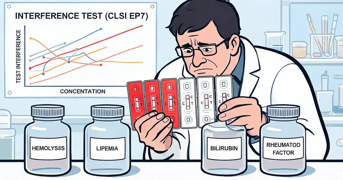
1. What Is Interference in a Lateral Flow Assay?
Interference in a lateral flow assay is any effect, other than the target analyte binding its antibody pair, that changes the intensity or interpretation of the test line or control line. It produces a false positive, a false negative, or a quantitative bias, and it is caused by substances that are either endogenous to the sample or introduced during collection.
It is useful to sort interference into four mechanistic classes, because each one calls for a different fix:
- Optical interference: colored or turbid substances — hemoglobin, bilirubin, lipids — absorb or scatter light in the readout window, masking a faint visual line or biasing a reflectance or fluorescence reader.
- Physical interference: viscosity and particulates change capillary flow rate and wetting, altering analyte–antibody contact time and background clearing.
- Chemical interference: redox-active and catalytic species (heme pseudo-peroxidase activity, ascorbate, glutathione) disturb label chemistry, most acutely in enzyme or chemiluminescence strips.
- Immunological interference: heterophilic antibodies, human anti-animal antibodies, rheumatoid factor, and the analyte itself in large excess (the hook effect) generate signal or suppress it through antibody-level cross-links.
The pitfalls below are ordered by how often they cause failed studies, starting with the interferent named in nearly every IVD specification: hemoglobin.
Critical Principle
A single interferent often acts through more than one mechanism at once. Hemoglobin is optical, chemical, and physical simultaneously. If you only correct the mechanism you can see — usually the color — the residual bias from the others will still fail a quantitative recovery study.
2. Pitfall 1: Treating Hemolysis as Only a Color Problem
Hemolysis is the most common endogenous interferent in immunoassay, and the frequent mistake is to assume that because a colloidal gold line is read by eye or reflectance, a red background is the only issue. Hemoglobin (Hb) — a 64.5 kDa tetramer carrying four iron-centered heme groups — interferes through three routes at once.
Optically, free Hb has a strong Soret absorption band near 410 nm plus Q-bands around 540 and 576 nm. On a visual gold strip this reddens the membrane and hides a faint line. On a fluorescent lateral flow (FIA) strip it is worse: the emission of many europium and fluorescent-latex labels falls inside the Hb absorption window, so part of the signal is absorbed before it reaches the detector — an inner filter effect. Chemiluminescence data show signal loss of 20–30% at 2 g/L Hb and 50–60% at 5 g/L.
Chemically, heme Fe2+ has pseudo-peroxidase activity that drives non-specific signal in any enzyme or luminescent chemistry, and oxidized heme from denatured Hb exposes a hydrophobic pocket that binds the antibody Fc region, the gold surface, and the nitrocellulose membrane — lifting background or stripping detector antibody off the conjugate. Physically and via matrix, hemolysis also releases potassium (raises ionic strength, weakens binding), glutathione and ascorbate (reduce label chemistry), and proteases (slowly degrade the capture antibody on the line).
| LFA label system | Readout peak | Hb sensitivity | Dominant mechanism |
|---|---|---|---|
| Colloidal gold (visual) | ~520–540 nm | Moderate | Color masking of faint line; heme non-specific binding |
| Fluorescent latex / europium (FIA) | ~530–615 nm | High | Inner filter effect at Soret band; reader bias |
| Enzyme / chemiluminescence strip | ~425–470 nm | Very high | Heme pseudo-peroxidase; spectral overlap |
| Latex agglutination (turbidimetric) | Scatter | Moderate | Turbidity and color offset |
Formulation-level mitigations: EDTA (5–10 mM) to chelate free iron, a mild reducing agent such as sodium sulfite to stop Hb oxidizing, and a radical scavenger such as propyl gallate for enzyme strips. Strengthen conjugate-pad and membrane blocking with casein plus BSA, raise running-buffer NaCl to 0.3–0.5 M and add Tween-20 to compete off hydrophobic binding, and buffer at pH 7.4–8.0, away from the pH 6–7 optimum of heme pseudo-peroxidase. For reader-based systems, a hemoglobin index channel (540 nm) that flags hemolyzed samples is the engineering backstop.
Lab Note
Test hemolysis with a mechanical lysate of washed red cells, not a commercial Hb powder in buffer. The powder lacks the potassium, LDH, and proteases behind much of the real-world bias, and will make the assay look more tolerant than it is.
3. Pitfall 2: Overlooking Lipemia, Icterus, and Turbidity
Hemolysis gets the attention; lipemia and icterus are the two interferents most often left out of an early panel and then discovered in field complaints.
Lipemia — elevated triglycerides and chylomicrons — acts physically and optically. Lipid particles slow and channel capillary flow, so the sample front reaches the test line later and less evenly, background clears incompletely, and a reflectance reader sees a hazy membrane. Very high triglycerides can also sequester lipophilic small-molecule analytes (some drugs, steroid hormones), lowering the free fraction that the assay detects.
Icterus — elevated bilirubin — is primarily optical for visual and reflectance strips: the yellow-brown background shifts the apparent line intensity and, for conjugated bilirubin, can add a weak chemical reducing effect. It is usually the easiest of the three classic interferents to design around but must still be quantified.
Practical handling:
- Build a filtering or asymmetric sample pad that retains lipid aggregates and particulates before the conjugate pad.
- Increase nonionic surfactant in the running buffer to improve wetting on a lipid-loaded membrane — but re-check conjugate stability, since the same change can destabilize gold. See surfactant selection mistakes in lateral flow assay development for the trade-offs.
- Where the analyte’s clinical decision range allows it, a 1:5 to 1:10 sample dilution in a well-designed running buffer is the simplest way to pull all three optical interferents below their threshold at once.
- Specify interferent limits in the instructions for use and, for reader-based systems, add an index channel or a turbidity check.
4. Pitfall 3: No High-Dose Hook Effect Check
The high-dose hook effect (prozone) is a false-negative or falsely low result that appears only at very high analyte concentrations. In a one-step sandwich strip, when analyte is in large molar excess it independently saturates the capture antibody on the test line and the detector antibody on the conjugate, so the two rarely meet on the same analyte molecule. Few labeled sandwiches form at the line, and it fades — sometimes to nothing — while the control line stays strong.
It is easy to miss because the standard analytical range used in early development never reaches the concentration where the hook begins. Analytes with a wide pathological range are the ones that bite: ferritin, hCG, CRP, procalcitonin, D-dimer, prolactin, and myoglobin.
"A one-step lateral flow assay that has never been challenged above its stated upper limit has not been tested for the hook effect — it has just been lucky with its samples."
How to design it out:
- Challenge deliberately. Run a dilution series at least 10–100× above the highest expected clinical value and confirm the line never drops below the positive threshold.
- Rebalance antibody mass. Increasing capture antibody on the line and detector antibody on the conjugate raises the concentration at which the hook begins.
- Consider a two-step or delayed-release format. A sequential flow (wash step, or a timed conjugate release pad) lets unbound analyte clear before the label arrives, largely removing the hook.
- Add a built-in dilution in the sample buffer for very wide-range analytes, accepting the sensitivity cost at the low end.
5. Pitfall 4: Skipping Heterophilic Antibody and Rheumatoid Factor Blocking
Immunological interference is the hardest failure to diagnose because the strip looks like it is working — it just reports the wrong answer. Heterophilic antibodies, human anti-animal antibodies (HAMA/HAAA), and rheumatoid factor (RF) are host immunoglobulins that bind animal IgG or the Fc region of other antibodies. On a sandwich strip they bridge the capture and detector antibodies directly, producing a false positive test line with no analyte present. RF is present in roughly 1–5% of the general population and far more in autoimmune patients, so a strip with no blocking will fail specificity on a normal donor panel.
The usual mistake is to leave blocking until a specificity problem shows up, then bolt on a single reagent. Blocking should be designed in and titrated:
| Blocking strategy | How it works | Where it goes |
|---|---|---|
| Non-immune animal IgG (mouse, and often goat/bovine) | Saturates heterophilic and anti-mouse binding sites before they reach the assay antibodies | Conjugate pad and/or sample pad |
| Polymerized or aggregated IgG / commercial heterophilic blocker | Multivalent decoy with high avidity for RF and heterophilic antibodies | Sample buffer |
| F(ab’)2 or scFv detector fragments | Removes the Fc target that RF and anti-Fc heterophilic antibodies recognize | Conjugate antibody design |
| Two-species antibody pair | Heterophilic antibody rarely bridges two unrelated host species at once | Pair selection |
A ready-made heterophilic blocking reagent is the fastest route for most projects, but the dose still has to be titrated against a defined heterophilic-positive and RF-positive sample panel — too little leaves residual false positives, too much can suppress a genuine low-end signal. Choosing the pair itself carefully upfront helps; see our guidance on IVD antibody screening pitfalls for how species and format choices interact with specificity.
6. Pitfall 5: Optimizing in Buffer Instead of the Real Clinical Matrix
Almost every interference pitfall above is invisible if the assay is only ever tested on analyte spiked into a clean buffer. The sample matrix is itself an interferent when it does not match what the assay will actually see.
- Whole blood vs serum vs plasma. Whole blood adds hematocrit-dependent viscosity and a red cell barrier at the sample pad; a strip tuned on serum will show a different flow time and line intensity on a 55% hematocrit sample. Anticoagulant matters too — EDTA, citrate, and heparin plasma are not interchangeable.
- Urine, saliva, and swab eluate vary widely in pH, ionic strength, and mucin or extraction-buffer content between donors; the running buffer has to normalize that range, not a single nominal sample.
- Biotin. For any strip built on a streptavidin–biotin capture system, patients taking high-dose biotin supplements can competitively block the interaction and cause a false negative (sandwich) or false positive (competitive). This is now an expected interference claim for biotin-based systems.
- Structural analogs. Endogenous metabolites, drug cross-reactants, and analyte degradation products should be screened against the pair — this is cross-reactivity, but presents in the field as interference.
The rule that prevents most of these: validate the assay in the same matrix, collection tube, and donor diversity that the finished product will encounter, and do it before the strip design is locked. Antibody pairs that already carry documented matrix and buffer compatibility data — like those on the Sekbio antibody development platform — shorten this loop considerably.
7. Building Anti-Interference In: A Development-Stage Framework
Interference tolerance is cheapest to build early and most expensive to retrofit. A workable sequence:
- Feasibility stage. List every interferent the target analyte’s published assays claim tolerance for, plus hemolysis, lipemia, and icterus by default. Decide the target claim levels now, because they drive antibody pair and format choice.
- Pair and format selection. Prefer recombinant antibodies with known epitopes, consider two host species, and decide one-step vs two-step based on the analyte’s clinical range and hook risk.
- Formulation screening. Titrate blockers (animal IgG, heterophilic blocker), chelators (EDTA), reducing agents, surfactant, and ionic strength against a spiked interferent panel — one variable at a time, re-checking conjugate stability after each change.
- Structured interference study. Run a CLSI EP7-style paired-difference design: test each interferent at two or more concentrations against control, at low and high analyte levels, and report bias as a percentage. This is the study that registration will expect.
- Lock the claim. State the validated tolerance limits (e.g. "no significant interference up to Hb 5 g/L, triglycerides 15 g/L, bilirubin 20 mg/dL") in the instructions for use.
- Lot QC. Re-qualify each new antibody, blocker, and surfactant lot against a reference interferent sample so tolerance does not drift silently in production.
For quantitative reader-based tests, add an engineering backstop: an optical index channel that flags hemolyzed, lipemic, or icteric samples, or a reference channel using non-immune IgG in place of the capture antibody so net signal subtracts matrix background.
8. Frequently Asked Questions — Lateral Flow Assay Interference
What is interference in a lateral flow assay?
Interference in a lateral flow assay is any effect, other than the analyte binding its antibody pair, that changes the test-line or control-line result. It includes optical interference (hemoglobin, bilirubin, or lipemia masking or scattering the signal), physical interference (viscosity and particulates altering capillary flow), chemical interference (heme pseudo-peroxidase activity and redox-active matrix components), and immunological interference (heterophilic antibodies, rheumatoid factor, and the high-dose hook effect). Each class needs a different mitigation, so identifying which one is acting is the first step.
How does hemolysis interfere with a lateral flow rapid test?
Hemolysis releases hemoglobin, which interferes in three ways. It colors the sample red, masking a faint visual test line and biasing reflectance or fluorescence readers. Its Soret absorption band near 410 nm overlaps the emission of many fluorescent labels, causing an inner filter effect that lowers signal. And exposed heme binds non-specifically to the colloidal gold conjugate and nitrocellulose membrane, raising background or suppressing the true line. Released potassium, LDH, and proteases add secondary matrix effects that a pure hemoglobin solution will not reproduce.
How long does an interference study take during rapid test development?
Formulation screening against a spiked interferent panel typically takes three to six weeks when run in parallel with antibody pair and conjugation work. The structured CLSI EP7 interference study that supports registration adds another two to four weeks, plus sample sourcing time for real hemolyzed, lipemic, icteric, and heterophilic-positive specimens. Teams that leave the whole effort until after design lock routinely add two to three months of rework.
What is the difference between the hook effect and cross-reactivity?
The high-dose hook effect is caused by the target analyte itself at an extremely high concentration, which saturates the capture and detector antibodies separately and collapses the test line, producing a false negative. Cross-reactivity is caused by a different molecule — a structural analog, metabolite, or degradation product — that the antibody pair also binds, usually producing a false positive or positive bias. Both are found by challenge testing, but the hook effect is checked with an analyte dilution series and cross-reactivity with a panel of related compounds.
Can I use a commercial hemoglobin powder to test hemolysis interference?
It is acceptable for a first-pass screen but not for the definitive study. A powder dissolved in buffer supplies hemoglobin without the potassium, lactate dehydrogenase, glutathione, and proteases that a real lysed red cell releases, so it underestimates the true bias. For the registration study, prepare a mechanical lysate from washed red cells and spike it into the intended matrix, following CLSI EP7 guidance on interferent preparation.
Does Sekbio offer antibody pairs validated for interference-prone assays?
Yes. Sekbio manufactures monoclonal and recombinant antibody pairs and antigens for lateral flow, ELISA, and CLIA platforms, with documented compatibility across common IVD buffer systems and matrix types, plus a dedicated heterophilic blocking reagent. If your team is troubleshooting an interference failure, our technical group can help identify whether the root cause is antibody chemistry, conjugation, or buffer formulation — see the antibody development platform for capabilities and data packages.
9. Summary
Anti-interference work in lateral flow assay development is a defined program, not a troubleshooting reaction:
- Hemolysis: treat it as optical, chemical, and physical at once — chelate iron, block hydrophobic sites, buffer above pH 7.4, and flag hemolyzed samples with an index channel.
- Lipemia and icterus: add them to the panel from the start; a filtering sample pad plus a modest sample dilution handles most cases.
- Hook effect: challenge every one-step assay well above its clinical range, rebalance antibody mass, or move to a two-step format.
- Heterophilic antibodies and rheumatoid factor: design blocking in — non-immune IgG, a heterophilic blocking reagent, Fc-free detector fragments, two-species pairs — and titrate against a positive panel.
- Matrix: validate in the real sample type, collection tube, and donor range, including biotin for streptavidin–biotin systems, before design lock.
- Framework: set claim levels at feasibility, screen formulation one variable at a time, run a CLSI EP7 study, state the limits in the IFU, and re-qualify lots.
Sekbio supplies monoclonal and recombinant antibody pairs for lateral flow, ELISA, and CLIA assays with matrix and buffer compatibility data, and a heterophilic blocking reagent for immunological interference. If an interference study is not going the way you expected, our technical team can help trace it to antibody, conjugate, or formulation.