IgM is the first antibody isotype to appear in an immune response and the largest immunoglobulin in human serum — a ~970 kDa pentamer built from five identical monomeric subunits joined by a J chain and interchain disulfide bonds. That size and quaternary structure make it an outstanding capture reagent for early-infection diagnostics and hemagglutination assays, but they also make IgM purification one of the least forgiving workflows in antibody development. Protocols that work perfectly for IgG routinely fail on IgM, and the failures are rarely obvious until an assay's sensitivity quietly collapses.
This guide walks through the mistakes that most often derail IgM purification — from reaching for a Protein A column out of habit, to over-concentrating a fragile pentamer until it aggregates, to skipping the one validation step that would have caught a dissociated pentamer before it reached the assay bench. Each section pairs the mistake with the fix IVD developers actually use in production.
1. What Is IgM Purification and Why Is It Different From IgG?
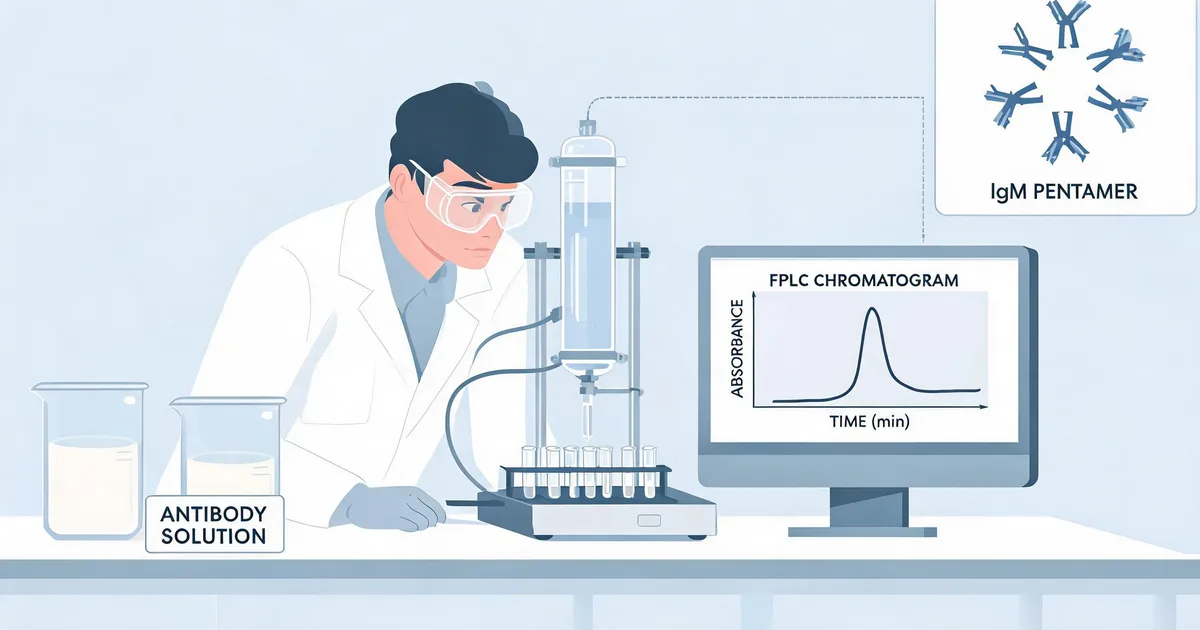
IgM purification is the process of isolating pentameric — or, without a functional J chain, hexameric — immunoglobulin M from serum, ascites fluid, or hybridoma culture supernatant while keeping its native quaternary structure intact. The goal is not just removing contaminating proteins; it is preserving a fragile, multi-subunit assembly that loses avidity and assay performance the moment it dissociates.
Four structural facts explain why IgM purification needs its own playbook rather than a scaled-up IgG protocol:
- Size and geometry: ~970 kDa with five Fab arms, versus ~150 kDa for a monomeric IgG — high avidity, but also high viscosity and aggregation propensity.
- Fc binding behavior: the mu heavy chain binds Protein A and Protein G with far lower, species-inconsistent affinity than IgG's Fc gamma region.
- Glycosylation and charge: heavily glycosylated and rich in sialic acid, giving IgM a strongly negative surface charge that changes how it behaves on ion-exchange resins.
- Bond sensitivity: held together by non-covalent contacts plus interchain disulfide bonds that are sensitive to pH extremes, reducing agents, and mechanical shear.
Because of these differences, IgM purification typically relies on capture chemistries that exploit these unique properties directly — thiophilic adsorption, PEG or euglobulin precipitation, or anti-mu chain affinity resins — rather than the Protein A/G workhorse used for IgG.
Definitional Note
An intact IgM pentamer, not total protein recovered, is what determines assay performance. Two purification lots can show identical protein concentration by A280 and behave completely differently in an agglutination or sandwich assay if one has dissociated to monomers.
2. Mistake 1: Reaching for Protein A/G Affinity Chromatography by Default
The single most common mistake in IgM purification is habit: developers who have purified dozens of IgG monoclonal antibodies default to a Protein A or Protein G column because it's the workflow they know. Protein A and Protein G bind the Fc gamma region with high, consistent affinity across most IgG subclasses — but the mu heavy chain of IgM interacts with these ligands weakly and inconsistently across species and IgM subclass, producing low, unpredictable recovery and, in some lots, no binding at all.
What to use instead:
- Thiophilic adsorption chromatography (T-gel type resins): binds IgM through a combined hydrophobic/electron-donor interaction under high-salt loading conditions, largely independent of Fc subclass, giving consistent recovery across species.
- Anti-mu chain affinity resin: an immunoaffinity approach using an antibody specific to the IgM heavy chain constant region, offering high specificity when cross-reactivity with other isotypes is a concern.
- PEG or euglobulin precipitation: a low-cost capture step that exploits IgM's large size and low solubility at reduced ionic strength, useful for first-pass concentration before a polishing chromatography step.
- Anion-exchange chromatography: IgM's dense sialylation gives it a strong negative charge at neutral-to-alkaline pH, making DEAE or Q-type resins effective for capture or polishing when paired with a shallow salt gradient.
Common Mistake
Testing a new IgM clone on a Protein A column and concluding the antibody "didn't express" is one of the more expensive false negatives in antibody development — the antibody is very likely present in the flow-through, not the eluate.
3. Mistake 2: Using the Wrong Buffer Ionic Strength for the Capture Step
Even when developers correctly move away from Protein A/G, the second most common error is applying the wrong loading buffer for the chosen capture chemistry. Thiophilic adsorption chromatography, unlike Protein A, requires a high-ionic-strength loading buffer — typically 0.5 M sodium sulfate or 0.8 M ammonium sulfate in phosphate buffer — to drive the hydrophobic/electron-donor interaction that binds IgM to the resin. Loading at physiological ionic strength, the default for most affinity workflows, produces little to no binding and a purification report that wrongly implicates the antibody rather than the buffer.
Anion-exchange capture runs into the opposite mistake: because IgM is so heavily sialylated, it binds anion-exchange resins even at moderate ionic strength, and low-ionic-strength loading buffers designed for typical serum proteins can co-bind a wide swath of contaminating glycoproteins, producing a "clean-looking" single peak that is anything but pure.
| Capture Method | Loading Buffer Strategy | Typical Recovery | Best Use Case |
|---|---|---|---|
| Protein A / G | Neutral pH, physiological ionic strength | <20%, inconsistent | Not recommended for IgM |
| Thiophilic (T-gel) | High ionic strength (0.5 M Na₂SO₃) | 60–85% | Universal capture, species-independent |
| Anti-mu affinity | Neutral pH, physiological ionic strength | 70–90% | High specificity, isotype discrimination |
| Anion exchange (DEAE/Q) | Low-to-moderate ionic strength, pH 7.5–8.5 | 50–75% | Polishing step after capture |
| PEG / euglobulin precipitation | Reduced ionic strength (precipitation, not chromatography) | 80–95% (crude) | Low-cost first-pass concentration |
Match the loading condition to the chemistry, not to habit, and run a small-scale buffer screen before committing an entire batch to the column.
4. Mistake 3: Ignoring Pentamer Stability During Elution and Buffer Exchange
A successful capture step can still produce a degraded product if the elution and buffer-exchange conditions stress the pentamer. IgM's five subunits are held together by non-covalent contacts and interchain disulfide bonds that are more sensitive to pH extremes, reducing agents, and mechanical shear than the disulfide framework of an IgG monomer.
Two conditions are frequent culprits:
- Low-pH glycine elution left unneutralized: acidic elution buffers common to affinity chromatography (pH 2.5–3.0) can drive partial or complete pentamer dissociation into monomeric or half-pentamer subunits if the eluate isn't neutralized to pH 7 within the collection tube immediately.
- Reducing agent carryover: trace beta-mercaptoethanol or DTT from upstream hybridoma culture supplements, or from shared buffer stocks, can reduce interchain disulfides and irreversibly dissociate the pentamer even at low concentrations.
"Pentamer integrity, not total protein recovered, is the number that actually predicts whether a purified IgM will perform in a sandwich or agglutination assay."
Buffer exchange itself should use gentle methods — dialysis or low-shear tangential flow filtration rather than repeated small-volume centrifugal spin concentration — and should avoid chelator-heavy formulations that can strip stabilizing divalent cations from the J-chain interface.
5. Mistake 4: Over-Concentrating IgM Until It Aggregates
IgM's large size and asymmetric five-armed geometry make it far more prone to concentration-dependent aggregation than IgG. Once total IgM concentration climbs above roughly 2–5 mg/mL, viscosity increases sharply, and localized supersaturation at a centrifugal filter membrane or ultrafiltration cassette can nucleate aggregates that never redissolve.
- Concentrate slowly, in stages, checking concentration and appearance — visible turbidity is an early aggregation signal — between spins rather than running one long centrifugation to a target volume.
- Avoid high g-force centrifugal spin concentrators with small membrane surface area; a lower g-force, larger-surface device reduces local concentration spikes at the membrane.
- Add a mild stabilizer — 2–5% sucrose or trehalose, or a modest NaCl adjustment — before the final concentration step, and confirm by SEC-HPLC that the stabilizer doesn't shift retention itself.
- Never vortex a concentrated IgM prep to mix; gentle pipetting or rocking is sufficient and avoids shear-induced aggregation.
Pro Tip
If your target application only needs 1–2 mg/mL working stock, stop concentrating there — pushing to a "round number" like 10 mg/mL for storage convenience is rarely worth the aggregation risk for IgM.
6. Mistake 5: Poor Storage Conditions and Skipping the Cryoglobulin Check
IgM's storage behavior is less forgiving than IgG's in two specific ways that developers routinely overlook.
First, repeated freeze-thaw cycles accelerate aggregation far more in IgM than in IgG — each cycle nucleates a small population of aggregates that accumulate irreversibly. Aliquoting into single-use volumes immediately after purification, rather than after the third or fourth freeze-thaw of a working stock, preserves far more usable pentamer over the product's shelf life.
Second, and more specific to IgM: a subset of IgM antibodies are cryoglobulins — they reversibly precipitate out of solution at reduced temperature, commonly around 4°C, and redissolve on warming. Running a cryoglobulin IgM through a cold-room chromatography column, or storing it at 4°C between purification steps, causes the antibody to precipitate directly on the resin bed or in the storage vial, producing what looks like catastrophic product loss but is actually a solubility artifact.
- Test a small aliquot of clarified sample at 4°C for 24–48 hours before committing to a cold-chain purification workflow.
- If cryoprecipitation is observed, keep all purification steps at room temperature and only introduce cold storage after confirming the final formulated product remains soluble at the intended storage temperature.
- Store long-term stock at -80°C with a cryoprotectant (sucrose or trehalose) rather than -20°C, where slower freezing promotes larger, more damaging ice crystal formation.
- Reserve 4°C storage for short-term working stock only, and confirm solubility at that temperature first.
Teams that outsource this step often use a partner's established antibody development and purification platform specifically because cryoglobulin screening and cold-chain validation are easy to skip under deadline pressure and expensive to discover late.
7. Mistake 6: Releasing Purified IgM Without Pentamer-Level Validation
The final and most consequential mistake is validating IgM purity the same way IgG is validated — with a single reducing SDS-PAGE gel. Reducing conditions break every disulfide bond in the sample, so a clean single band at approximately 70–75 kDa (the reduced mu heavy chain) confirms the protein sequence looks right but says nothing about whether the pentamer was still intact before the gel was run. A batch that dissociated to monomers during elution can produce an identical reducing gel to a batch with perfect pentamer integrity.
A validation panel that actually distinguishes these two batches needs at least three orthogonal methods:
| Method | What It Confirms | What It Misses |
|---|---|---|
| Reducing SDS-PAGE | Heavy/light chain identity and purity | Pentamer vs. monomer state (destroyed by reduction) |
| Non-reducing / native PAGE | High-MW complex presence, gross degradation | Precise pentamer:aggregate ratio |
| SEC-HPLC | Quantitative pentamer:monomer:aggregate ratio | Amino acid-level identity |
| A280/A320 turbidity ratio | Soluble aggregate / turbidity screen | Low-level oligomeric species |
Running SEC-HPLC on every purification lot, not just during method development, is the single highest-value quality control step for IgM — it is the only method in the panel that directly reports the percentage of intact pentamer, the number that correlates with agglutination titer and sandwich-assay signal. For IVD developers pairing purified IgM into a sandwich immunoassay format, pentamer purity below roughly 85–90% by SEC-HPLC area is a strong predictor of downstream sensitivity problems, even when the reducing gel looks flawless.
8. Frequently Asked Questions — IgM Purification
What is IgM purification?
IgM purification is the process of isolating pentameric immunoglobulin M from serum, ascites fluid, or hybridoma culture supernatant while preserving its native five-subunit quaternary structure. Because the pentamer's avidity depends on its intact assembly, successful IgM purification protocols are optimized around structural preservation, not just removing contaminating proteins the way IgG purification is.
How long does IgM purification take?
A single-lot IgM purification, from clarified sample to validated, formulated product, typically takes 3–5 working days using a pre-optimized thiophilic or anion-exchange capture step followed by SEC polishing and SEC-HPLC validation. Method development for a novel IgM clone — including a buffer and resin screen, cryoglobulin testing, and stability validation — usually adds 1–2 additional weeks before the production protocol is finalized.
Can I use Protein A or Protein G to purify IgM antibodies?
Not reliably. Protein A and Protein G bind the Fc gamma region of IgG with high, consistent affinity, but the mu heavy chain of IgM interacts with both ligands weakly and inconsistently across species and IgM subclass. Recovery from a Protein A or Protein G column is typically low and unpredictable, and some IgM clones show essentially no binding at all. Thiophilic adsorption chromatography, anti-mu chain affinity resin, or anion-exchange chromatography are the standard alternatives.
What is the difference between thiophilic chromatography and ion-exchange chromatography for IgM?
Thiophilic (T-gel) chromatography captures IgM through a combined hydrophobic and electron-donor interaction under high-ionic-strength loading conditions, largely independent of antibody subclass or species, making it a reliable universal capture step. Anion-exchange chromatography separates IgM based on its strongly negative charge from dense sialylation and is more commonly used as a polishing step after capture, since it can co-bind other highly sialylated serum glycoproteins if ionic strength isn't tightly controlled.
How do you validate IgM pentamer integrity after purification?
Pentamer integrity should be validated with SEC-HPLC, which quantifies the ratio of intact pentamer to dissociated monomer and higher-order aggregate in a single run, supplemented with non-reducing or native PAGE to confirm the high-molecular-weight complex is present. A reducing SDS-PAGE gel alone is not sufficient — reduction breaks the interchain disulfide bonds holding the pentamer together, so it cannot distinguish an intact pentamer from a batch that already dissociated during purification.
Does Sekbio offer custom IgM antibody purification and characterization services?
Yes. Sekbio develops and validates purification protocols for IgM and other complex immunoglobulin formats, including capture chemistry selection, cryoglobulin screening, and SEC-HPLC-based pentamer integrity validation, alongside our monoclonal antibody pairing and recombinant antigen development work. Visit our antibody development and purification platform page to discuss your IgM project.
9. Summary
Building a reliable IgM purification workflow comes down to six recurring fixes:
- Skip Protein A/G by default: use thiophilic adsorption, anti-mu affinity, PEG/euglobulin precipitation, or anion exchange instead.
- Match ionic strength to chemistry: high salt for thiophilic capture, controlled low-to-moderate salt for anion exchange.
- Protect pentamer integrity at elution: neutralize acidic eluate immediately and eliminate reducing-agent carryover.
- Concentrate gradually: stop at the concentration your assay actually needs and monitor for early turbidity.
- Test for cryoglobulin behavior before cold-chain purification: a 4°C solubility check takes a day and prevents catastrophic column loss.
- Validate with SEC-HPLC, not just reducing SDS-PAGE: pentamer:monomer:aggregate ratio is what predicts assay performance.
At Sekbio, we develop and manufacture recombinant antigens and monoclonal antibodies — including specialized IgM- and IgE-class capture reagents — for IVD assay developers building sandwich immunoassays, agglutination tests, and rapid diagnostics. Our validated antibody pair catalog and custom purification services are backed by SEC-HPLC-confirmed pentamer integrity data. If you're purifying or sourcing IgM reagents, let's talk.