Progesterone assay development runs into a problem most protein biomarkers never face: the target is a small steroid molecule structurally almost identical to another clinically important hormone circulating in the same sample. Antibody pair selection for progesterone isn't primarily about finding two non-overlapping epitopes — that option doesn't exist at 314 Da. It's about selecting a single antibody with enough affinity and enough discrimination against its closest structural neighbor to survive real patient samples.
This guide is written for IVD developers designing a progesterone competitive assay from the antibody up: why the format choice is fixed by molecular size, how to validate against the specific cross-reactant that causes the most false elevations, and what changes when the same antibody needs to work across both a CLIA analyzer and a point-of-care LFA cartridge.
1. What Is Progesterone Antibody Pair Design?
Progesterone antibody pair design is the process of selecting and validating a single monoclonal antibody — plus its labelled tracer counterpart — for use in a competitive immunoassay, optimizing for affinity, cross-reactivity exclusion, and compatibility with the intended CLIA and/or LFA platform. This is a fundamentally different design exercise from sandwich-format antibody pairing used for larger protein biomarkers, where two antibodies must be selected to bind non-overlapping epitopes simultaneously.
For clinical decision cutoffs and normal reference ranges by cycle phase and pregnancy stage, see our progesterone normal range guide. This article covers the reagent design problem behind those numbers — how the antibody itself has to be built to hit them reliably.
2. Why Competitive Format Is Mandatory
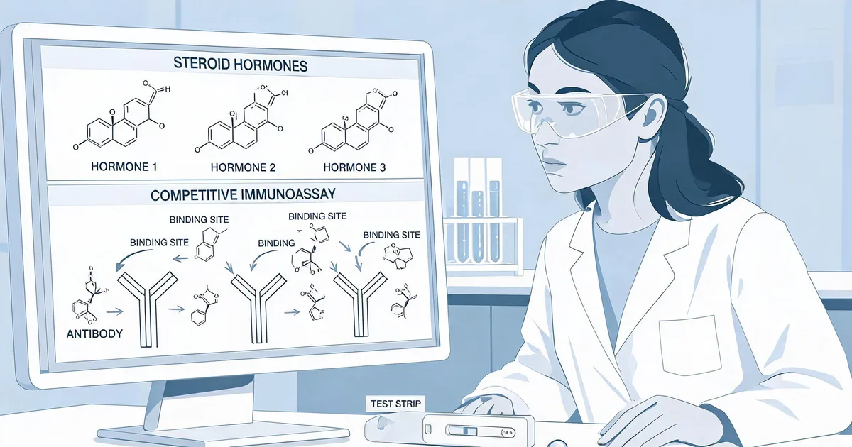
Progesterone (MW 314 Da) is far too small to present two spatially distinct, simultaneously accessible epitopes to two separate IgG antibodies. Every progesterone immunoassay — CLIA, ELISA, or LFA — therefore uses a competitive format: unlabelled sample progesterone and a fixed quantity of labelled progesterone tracer compete for a limited pool of antibody binding sites, and signal is inversely proportional to sample concentration.
This constraint shapes antibody selection criteria that don't apply to sandwich-format development. The antibody needs an affinity constant in a fairly narrow window: too low, and the competition reaction is too weak to produce a usable signal gradient across the clinical range; too high, and the tracer can't be efficiently displaced by physiological sample concentrations, compressing the assay's sensitivity at the low end. Hapten-carrier conjugate design during antibody generation — how progesterone is chemically linked to a carrier protein to raise an immune response in the first place — also determines which face of the steroid molecule remains exposed for antibody recognition, which in turn determines the cross-reactivity profile discussed next.
Critical Principle
Because competitive format has no second antibody to add specificity, all of the selectivity burden sits on a single binding site. This makes cross-reactivity screening, not epitope mapping, the central validation exercise for progesterone antibody selection.
3. The 17-Hydroxyprogesterone Cross-Reactivity Problem
Progesterone shares its steroid backbone with several circulating hormones, but one cross-reactant matters more than the rest: 17-hydroxyprogesterone (17-OHP). It differs from progesterone by a single hydroxyl group at the C17 position — the smallest structural difference between progesterone and any other clinically relevant steroid — which makes it the hardest cross-reactant to exclude through antibody selectivity alone.
The clinical stakes are higher than a generic false-positive risk. 17-OHP is itself the primary biomarker for congenital adrenal hyperplasia (CAH), and CAH patients — including newborns on CAH screening panels — can have 17-OHP concentrations dramatically elevated above normal. An insufficiently selective progesterone antibody in this population doesn't produce a modest bias; it can report a falsely elevated progesterone result driven almost entirely by 17-OHP cross-reaction, which is clinically misleading in exactly the population where accurate steroid panel results matter most.
| Cross-Reactant | Structural Relationship | Target Cross-Reactivity |
|---|---|---|
| 17-Hydroxyprogesterone | Single C17 hydroxyl difference | <1–2% |
| Pregnenolone | Precursor steroid, similar backbone | <2% |
| Corticosterone | Related adrenal steroid | <1% |
Validating against this threshold requires spiking recovery studies across the full CAH-relevant concentration range, not a single reference-range data point — cross-reactivity is not always linear, and an antibody that looks acceptably selective at normal 17-OHP concentrations can behave differently at the markedly elevated concentrations seen in CAH patients.
"For progesterone, the antibody selection question isn't 'does it bind progesterone well' — every reasonable candidate does. It's 'does it still ignore 17-hydroxyprogesterone at the concentrations where that distinction actually matters clinically.'"
4. Matrix Effects: Serum vs. Whole Blood in LFA
Progesterone circulates highly protein-bound, primarily to albumin and corticosteroid-binding globulin, with only a small free fraction available to react in an immunoassay. A competitive assay validated on serum — where the sample has already been separated from cellular components — can behave differently when ported directly to a whole blood lateral flow cartridge without matrix-matched revalidation.
- Hematocrit-driven separation efficiency: LFA plasma-separation membranes must consistently deliver the fraction of the sample containing progesterone to the reaction zone regardless of a patient's hematocrit, which varies more between individuals than most developers initially budget for.
- Residual protein binding: even after plasma separation, the same protein-binding equilibrium that governs serum behavior is present in whole blood plasma, and the tracer competition kinetics must be validated against this matrix directly rather than assumed from serum data.
- Revalidation, not extrapolation: the safest development path treats whole blood LFA as a separate validation exercise from serum CLIA, using matrix-matched spiked samples rather than applying a serum-derived correction factor.
5. CLIA + LFA Dual-Format Development Workflow
Developing both a CLIA analyzer reagent and an LFA point-of-care cartridge from the same antibody source is common in fertility diagnostics, where centralized labs and clinic-based point-of-care testing serve different parts of the same care pathway. A single antibody can often support both, provided it is available in two preparation states:
- Unconjugated, purified antibody for CLIA use as the solid-phase capture reagent, paired with a labelled progesterone tracer (enzyme or chemiluminescent label) in the analyzer's reaction chemistry.
- Conjugation-ready antibody for LFA labelling, typically to colloidal gold or a fluorescent particle, striped onto the test line for competitive lateral flow detection.
Qualifying a single antibody source across both formats reduces the cross-reactivity and specificity validation burden to one campaign instead of two, since the binding-site selectivity characterized for CLIA carries over directly to the LFA format — only the matrix and detection chemistry change, not the fundamental antibody-antigen interaction being exploited.
IVD Application Note
Sekbio's progesterone antibody (S01-Prog-1S) is characterized for both competitive CLIA and LFA use, purity >90% by SDS-PAGE with inter-batch CV <10%, supporting single-source qualification across dual-format development programs.
6. Calibration & Dynamic Range Design
Progesterone's clinical concentration range spans roughly three orders of magnitude — from sub-3 nmol/L follicular-phase baselines to over 500 nmol/L during IVF luteal support — which is unusually wide for a single competitive assay to cover without dilution. Calibration curve design has to account for this full span while preserving resolution at the clinically critical low end, where the 6 nmol/L ectopic/miscarriage-risk threshold and the 25 nmol/L early-pregnancy-viability threshold sit close together.
- Non-linear calibration modeling (4-parameter or 5-parameter logistic curves) is standard for competitive assays given the inherently non-linear dose-response relationship, and calibrator spacing should be denser in the 3–30 nmol/L clinical decision zone than at the high end of the range.
- High-dose hook effect is not a concern the way it is for sandwich assays, since competitive format signal decreases monotonically with concentration rather than following a bell curve — but confirming true monotonicity across the full validated range remains part of standard characterization.
7. Assay & Quality Considerations
Beyond antibody selection itself, a few manufacturing and QC factors determine whether a progesterone competitive assay performs consistently in production:
- Lot-to-lot affinity consistency: because assay sensitivity is directly tied to the antibody's affinity constant, inter-batch CV needs tight control (<10%) to avoid calibration drift between reagent lots reaching the field.
- Tracer stability: the labelled progesterone tracer degrades independently of the antibody and needs its own shelf-life validation, since tracer degradation shifts the competition equilibrium in ways that mimic antibody performance loss.
- Cross-reactivity re-verification per lot: because selectivity against 17-OHP is the single most clinically important performance characteristic, spot-checking cross-reactivity on a subset of production lots — not just the original development lot — protects against drift introduced by manufacturing process changes.
8. Summary
Progesterone antibody pair design is shaped by the molecule's small size and its close structural relatives:
- Format: competitive immunoassay only — sandwich format is not physically possible at 314 Da.
- Cross-reactivity: 17-hydroxyprogesterone is the critical validation target, with clinical stakes tied to CAH testing accuracy; target <1–2% cross-reactivity verified across the full clinical concentration range.
- Matrix: whole blood LFA requires its own matrix-matched validation rather than extrapolation from serum CLIA data.
- Dual-format development: a single well-characterized antibody can often serve both CLIA and LFA, reducing validation burden.
- Dynamic range: non-linear calibration modeling with denser calibrator spacing in the 3–30 nmol/L clinical decision zone.
At Sekbio, we manufacture the competitive-format progesterone antibody behind both CLIA and LFA fertility assay development, characterized for the cross-reactivity exclusion this class of assay demands. If you're building a progesterone panel — standalone or alongside AMH and other fertility markers — our team can walk through cross-reactivity and lot-consistency data for your platform.
Frequently Asked Questions — Progesterone Antibody Design
Why can't progesterone be measured with a sandwich immunoassay?
Progesterone is a 314 Da steroid molecule with a single accessible epitope, too small to accommodate two antibodies binding simultaneously at spatially distinct sites the way a sandwich format requires. Competitive immunoassay is therefore the only viable format: sample progesterone and a labelled progesterone tracer compete for a fixed number of antibody binding sites, and signal is inversely proportional to sample concentration.
Why is 17-hydroxyprogesterone the biggest cross-reactivity risk for progesterone assays?
17-OH-progesterone differs from progesterone by a single hydroxyl group at the C17 position, making it the most structurally similar circulating steroid and the hardest cross-reactant to exclude through antibody selectivity alone. Because 17-OH-progesterone is itself an important marker for congenital adrenal hyperplasia, patients with CAH can have markedly elevated 17-OH-progesterone that falsely inflates an insufficiently selective progesterone assay result.
What cross-reactivity threshold should a progesterone antibody meet against 17-OH-progesterone?
Clinical-grade progesterone antibodies should demonstrate cross-reactivity below roughly 1-2% against 17-OH-progesterone at physiological concentrations, verified by spiking recovery studies across the CAH-relevant concentration range rather than a single reference-range data point, since cross-reactivity is not always linear across a compound's full clinical concentration range.
Why does whole blood matter differently than serum for progesterone LFA design?
Progesterone is highly protein-bound in serum, primarily to albumin and corticosteroid-binding globulin, and a whole blood lateral flow format must account for both the hematocrit-driven plasma separation efficiency and any residual protein binding that could reduce the free tracer competition available to react with the capture antibody. Assays validated only on serum can show a systematic bias when ported directly to a whole blood cartridge without matrix-matched revalidation.
Can the same progesterone antibody be used for both CLIA and LFA development?
Often yes, provided the antibody's affinity constant falls in the right range for both formats and it is available unconjugated for CLIA capture use and as a conjugation-ready format for LFA labelling. Sekbio's S01-Prog-1S is characterized for both applications, letting a developer qualify a single antibody source across a benchtop CLIA analyzer and a point-of-care LFA cartridge.
Does Sekbio supply progesterone antibodies for OEM assay development?
Yes. Sekbio supplies S01-Prog-1S, a monoclonal antibody characterized for competitive CLIA and LFA progesterone immunoassay development, purity >90% by SDS-PAGE with inter-batch CV <10%. Explore the datasheet or discuss cross-reactivity validation data through our antibody development services.