Most AMH content online covers what the hormone means for a patient — normal ranges by age, PCOS thresholds, IVF prognosis. Almost none of it addresses the question an IVD developer actually needs answered: which antibody pair should you use, and why do different AMH assays disagree with each other even when testing the same sample?
The answer lies in AMH's biology, not just its clinical interpretation. This guide walks through the antibody pair selection process — form specificity, epitope pairing, cross-reactivity screening, and dynamic range validation — that determines whether an AMH assay produces results clinicians can trust.
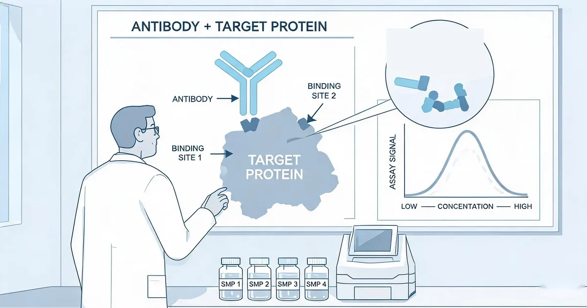
1. What Is AMH Form Specificity, and Why Does It Matter?
Anti-Müllerian hormone is not secreted in a single, uniform molecular form. Granulosa cells release it first as an inactive precursor, proAMH, which is then proteolytically cleaved into an N-terminal and a C-terminal fragment. These two fragments typically remain non-covalently associated as the AMHN,C complex, but circulating blood contains a mixture of proAMH and the cleaved complex in proportions that vary between individuals and even between menstrual cycle phases.
This matters because antibody pairs differ in which form they recognize. A capture/detection pair raised against epitopes on the cleaved complex may under-detect uncleaved proAMH, and vice versa. This is the single biggest reason AMH results can differ meaningfully between assay platforms measuring the same patient sample — it is an antibody specificity issue, not simply a calibration difference.
Critical Principle
Before screening any antibody clones, decide explicitly whether your assay is intended to measure total AMH (both forms) or is being developed for a specific research application targeting one form selectively. This decision determines every subsequent step of pair selection.
2. Step 1: Define Which AMH Form Your Assay Must Detect
Nearly all commercial fertility AMH assays are designed to measure total AMH, since clinical decision-making (ovarian reserve assessment, IVF protocol selection, PCOS diagnosis) relies on total circulating hormone rather than the proAMH/cleaved-complex ratio specifically.
- Total AMH assays typically pair a capture antibody against a conserved region present in both proAMH and the cleaved complex with a detection antibody against a second conserved epitope, maximizing recognition regardless of processing state.
- Form-specific assays (used mainly in research rather than routine clinical fertility testing) deliberately target epitopes unique to proAMH or to the cleaved complex, and require reference standards for each form separately.
For OEM fertility IVD development, defining "total AMH" as the target from the outset avoids a costly late-stage discovery that your antibody pair has been systematically under-detecting one circulating form.
3. Step 2: Screen Capture & Detection Epitope Pairs
Once the target form is defined, candidate antibody clones are screened in a checkerboard matrix to identify pairs that bind non-overlapping epitopes and form a stable sandwich complex:
- Test every capture-detection combination across a titration matrix against purified or recombinant AMH standard.
- Confirm each pair produces a dose-dependent signal with low background in a "no antigen" control well.
- Select the pair combination with the highest signal-to-noise ratio, prioritizing consistent performance across the full form-mixture (proAMH + cleaved complex) rather than raw peak signal against a single purified form.
"An antibody pair that performs beautifully against purified recombinant AMH standard but poorly against pooled clinical serum is almost always a form-specificity mismatch, not a sensitivity problem."
4. Step 3: Rule Out Cross-Reactivity With Inhibin B
AMH belongs to the TGF-β superfamily, which also includes inhibin B, activin, and other structurally related reproductive hormones — several of which are measured in the same fertility panels as AMH. Cross-reactivity between an AMH antibody pair and these related proteins can produce falsely elevated results, particularly in patients where inhibin B is also elevated.
- Screen at high concentration: test candidate antibody pairs against inhibin B and other TGF-β family members at concentrations well above their normal physiological range (typically 100× the target analyte's upper reference limit) to expose any low-affinity cross-reactivity that would be invisible at normal concentrations.
- Screen against structurally similar reproductive hormones routinely co-panelled with AMH, since a fertility test panel combining multiple hormones amplifies the clinical consequence of any single cross-reactive result.
- Document specificity data as part of the validation file — regulatory reviewers for fertility IVD panels routinely request cross-reactivity data against structurally related hormones.
5. Step 4: Validate Dynamic Range for DOR to PCOS
AMH's clinical range spans roughly two orders of magnitude in a single patient population — from <1 ng/mL in diminished ovarian reserve (DOR) and premature ovarian insufficiency, through normal reserve, up to >15–20 ng/mL in polycystic ovary syndrome, where AMH is characteristically elevated 2–4× above normal. A single antibody pair must remain accurate across this entire spread without requiring sample dilution for most clinical presentations.
- Low-end sensitivity: distinguishing DOR from low-normal reserve requires a low limit of detection and blank limit, since the clinically important decisions cluster below 1 ng/mL.
- High-end linearity and hook-effect resistance: at PCOS-range concentrations, verify the antibody pair does not exhibit a hook (prozone) effect — where antigen excess saturates both capture and detection antibodies and produces a falsely low reading — by testing serial dilutions of high-concentration samples and confirming proportional recovery.
- Two-step incubation: a wash step between sample addition and detection antibody addition is standard practice for high-range antigens like PCOS-level AMH, since it prevents simultaneous saturation of both antibodies.
6. Step 5: Choose FIA/POCT or CLIA Format
Format choice depends on where the assay will be deployed and what turnaround time the clinical workflow requires:
| Format | Best Setting | Key Requirement |
|---|---|---|
| FIA / Lateral Flow POCT | Fertility clinics, near-patient testing | Dry-format conjugate stability; fast turnaround (15–30 min) |
| CLIA | Centralized reference labs | Lowest LoD for DOR discrimination; automated high-throughput workflow |
The same core capture/detection clone pair can often serve both formats, but conjugation chemistry differs substantially — lateral flow requires colloidal gold or fluorescent label conjugation with dried-reagent stability, while CLIA requires chemiluminescent or enzyme labels optimized for a wet, automated platform. Each format requires independent validation even when reusing the same underlying antibody clones.
IVD Application Note
Sekbio's AMH antibody pair is built on a validated FIA sandwich format covering 0.1–20 ng/mL with LoD of 0.1 ng/mL and within-run CV ≤6.6%, benchmarked against Roche Elecsys across 60 clinical samples spanning the DOR-to-PCOS range.
7. Lessons From the Gen II AMH Assay Variability Episode
The clearest real-world illustration of why antibody pair selection matters is the widely documented variability seen with early-generation automated AMH assays, where complement interference and sample-handling sensitivity caused significant under-recovery in some serum samples, prompting revised pretreatment protocols before the issue was resolved. Independent method-comparison studies published in the years following raised the issue prominently enough that it reshaped how the field validates new AMH assays.
The lasting lesson for antibody pair selection: analytical performance against purified calibrator material is necessary but not sufficient. A pair must also be challenged against real clinical serum or plasma across diverse patient populations, since matrix effects and sample-handling sensitivity that never appear in calibrator-only testing can still compromise clinical accuracy. For further reading on AMH form biology and assay comparison methodology, see proAMH/AMHN,C complex studies on PubMed and AMH assay variability literature on PubMed.
8. Summary
Selecting an AMH antibody pair for fertility IVD comes down to five checks:
- Form specificity: confirm the pair recognizes both proAMH and the cleaved AMHN,C complex if a total-AMH result is the goal.
- Epitope pairing: validate non-overlapping capture/detection binding via checkerboard titration against clinical serum, not just purified standard.
- Cross-reactivity: screen against inhibin B and other TGF-β family members at high concentration.
- Dynamic range: confirm linearity and hook-effect resistance from <1 ng/mL (DOR) through >15–20 ng/mL (PCOS) without dilution.
- Format fit: match conjugation chemistry to FIA/POCT or CLIA deployment, validating each format independently.
At Sekbio, we manufacture the monoclonal antibody pairs behind fertility hormone panels, validated against reference platforms across the full clinical concentration range. If you're developing an AMH assay and need reagent-level performance and cross-reactivity data for your validation file, our team can walk through the datasheet with you.
Pair selection is only half of a registration file. Pudite obtained a product registration certificate for its AMH assay built on Sekbio antibodies; our IVDR 2026 antibody supplier documentation guide covers the CoA, lot consistency and stability data a reviewer will expect for the antibody pair.
Frequently Asked Questions — AMH Antibody Pair Selection
What is the difference between proAMH and the cleaved AMHN,C complex?
AMH is secreted as an inactive precursor, proAMH, which is proteolytically cleaved into an N-terminal and C-terminal fragment that remain non-covalently associated as the AMHN,C complex. Most circulating AMH exists as a mixture of both forms, and different antibody pairs preferentially recognize one form over the other, which is a major source of inter-assay variability when comparing results across platforms.
How long does AMH antibody pair validation typically take for OEM development?
A full validation cycle covering epitope pairing, cross-reactivity screening, and clinical method comparison typically takes 8–12 weeks, depending on how many candidate antibody clones are being screened and whether a reference-method comparison study (such as against Roche Elecsys) is included.
Can the same AMH antibody pair be used for both FIA POCT and CLIA formats?
The same core capture/detection antibody pair can often be adapted to both formats, but the conjugation chemistry and stabilization requirements differ substantially — FIA lateral flow needs the pair conjugated to a fluorescent or colloidal gold label with dry-format stability, while CLIA needs a chemiluminescent or enzyme label optimized for a wet, automated analyzer workflow. Cross-format validation is required even when reusing the same antibody clones.
What is the difference between AMH and inhibin B for ovarian reserve testing?
Both are TGF-β superfamily hormones produced by ovarian granulosa cells, but AMH is largely cycle-independent and reflects the pre-antral/small antral follicle pool, making it the preferred single measurement for ovarian reserve. Inhibin B fluctuates across the menstrual cycle and is a less reliable stand-alone marker, though its structural similarity to AMH makes cross-reactivity screening between the two an essential antibody pair validation step.
How do you validate an AMH antibody pair against high PCOS-range concentrations?
Test the antibody pair against calibrated samples spanning the full clinical range, including concentrations above 15–20 ng/mL typical of PCOS, and confirm the dose-response curve remains linear at the upper range without a hook effect (a false low reading caused by antigen excess saturating both capture and detection antibodies) using a two-step incubation protocol with an intervening wash.
Does Sekbio offer an AMH antibody pair for OEM assay development?
Yes. Sekbio's AMH antibody pair is validated in FIA sandwich format across a 0.1–20 ng/mL linear range with CV ≤6.6%, benchmarked against Roche Elecsys across 60 clinical samples. Explore our full antibody development services for custom fertility panel projects.