Many IVD teams treat surfactant selection as an afterthought — reaching for 0.05% Tween-20 because "that's what the last project used" — only to discover late in development that the running buffer destabilizes the gold conjugate, or that the striping buffer is quietly suppressing antibody immobilization on the nitrocellulose membrane. Surfactant choice touches nearly every component of a lateral flow strip, and a mismatch in any one of them can produce a symptom that looks exactly like an antibody problem.
This guide walks through the surfactant selection mistakes we see most often in lateral flow immunoassay (LFA) development, why each happens, and how to replace ad hoc buffer copying with a systematic screening process.
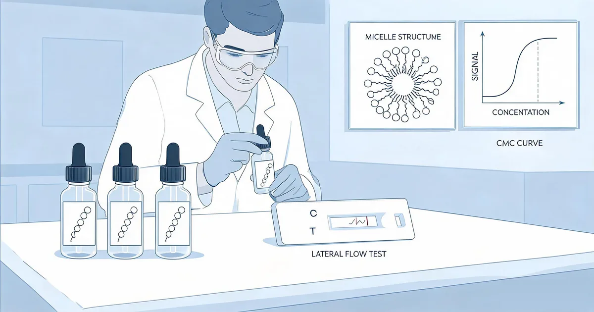
1. What Is the Role of Surfactants in Lateral Flow Immunoassay Development?
A surfactant (surface-active agent) in lateral flow immunoassay development is a molecule added to the running buffer, conjugate pad, sample pad, or blocking/striping buffer to reduce surface tension, control the wetting and capillary flow characteristics of the nitrocellulose membrane, minimize non-specific protein binding from the sample matrix, and stabilize colloidal gold or latex particle conjugates in suspension.
In practice, a surfactant is doing several distinct jobs at once, and a formulation that works well for one job can actively work against another:
- Wetting agent: lowers surface tension so sample and buffer flow evenly and quickly across the membrane instead of pooling or channeling.
- Flow modifier: influences capillary migration speed, which affects how long the analyte has to bind the capture antibody at the test line.
- Conjugate stabilizer: keeps colloidal gold or latex particles from aggregating in the conjugate pad and running buffer.
- Non-specific binding blocker: reduces matrix proteins and membrane surface sites from generating background signal.
The vast majority of validated LFA formulations use nonionic surfactants — polysorbate 20 (Tween-20), Triton X-100, and Pluronic F-68 (Poloxamer 188) are the most common. Ionic surfactants are largely avoided: anionic surfactants like SDS denature antibody tertiary structure, and cationic surfactants can be electrostatically attracted to the negatively charged nitrocellulose membrane surface.
Critical Principle
Surfactant behavior is interdependent with every other buffer component. Changing the running buffer surfactant concentration can shift capillary flow time, which changes analyte-antibody contact time, which changes assay sensitivity. Optimize systematically, one variable at a time, and re-validate downstream steps after any change.
2. Mistake 1: Choosing Surfactant Type by Habit Instead of HLB Value and Charge Compatibility
The single most common mistake is defaulting to whatever surfactant the last project used, without checking whether its Hydrophile-Lipophile Balance (HLB) and charge properties actually suit the current antibody pair and membrane system. HLB describes how hydrophilic or lipophilic a surfactant is — it determines how strongly the molecule interacts with hydrophobic protein patches versus staying dissolved in the aqueous phase.
2.1 Common LFA Surfactants Compared
| Surfactant | Type | Approx. HLB | Typical LFA Use |
|---|---|---|---|
| Tween-20 (Polysorbate 20) | Nonionic | ~16.7 | Running buffer, sample diluent, general blocking |
| Triton X-100 | Nonionic | ~13.5 | Membrane pre-treatment, viral lysis buffers |
| Pluronic F-68 (Poloxamer 188) | Nonionic | ~29 | Whole blood matrices, low-foam formulations |
| SDS | Anionic | Ionic (no HLB) | Avoid — denatures antibody tertiary structure |
| CTAB | Cationic | Ionic (no HLB) | Avoid — disrupts nitrocellulose surface charge |
Because nitrocellulose membranes carry a net negative surface charge, cationic surfactants can be electrostatically drawn to the membrane and interfere with protein immobilization or cause streaking. Anionic surfactants such as SDS denature antibody structure at even low concentrations, destroying binding activity outright.
Common Mistake
Copying a surfactant blend from a generic datasheet without checking the antibody pair's isoelectric point and hydrophobic patch distribution. Hydrophobic antibodies can lose activity even in nonionic surfactants once micelle formation disrupts the same interactions that stabilize the antibody's fold.
3. Mistake 2: Setting Concentration Without Reference to the Critical Micelle Concentration (CMC)
The critical micelle concentration (CMC) is the concentration above which surfactant molecules self-assemble into micelles rather than remaining as free monomers at the interface. Below the CMC, surface tension keeps dropping as concentration increases; above it, surface tension plateaus and additional surfactant does little more for wetting — but continues to affect protein-protein and protein-membrane interactions.
Tween-20 has a CMC of approximately 0.06 mM (~0.0074% w/v); Triton X-100 is approximately 0.22–0.24 mM (~0.014–0.016% w/v). Most validated LFA running buffers use Tween-20 in the 0.05–0.5% v/v range — roughly 7 to 70 times the CMC — by design, to guarantee saturation wetting even after some surfactant is consumed by adsorption to the plastic cassette housing and membrane fibers.
The mistake is copying a concentration from a paper or supplier datasheet without titrating it against your own membrane, conjugate, and clinical matrix. Too little surfactant produces incomplete wetting, streaking, and elevated background from non-specific matrix binding. Too much accelerates capillary migration past the point where the analyte has time to bind the capture antibody, reducing signal intensity, and at high enough levels can strip loosely bound antibody off the conjugate surface entirely.
"Surfactant concentration in a lateral flow assay isn't a number to copy from a paper — it's a titration to run against your own membrane, conjugate, and clinical matrix."
4. Mistake 3: Adding Surfactant to the Conjugate Pad Without Verifying Colloidal Gold/Latex Conjugate Stability
Teams frequently optimize running buffer surfactant concentration using only nitrocellulose flow criteria, then discover that when the same buffer rehydrates the conjugate pad, it strips loosely adsorbed antibody off the gold nanoparticle surface — causing aggregation and a loss of detection signal that looks like an antibody pairing failure.
Running buffer optimization and conjugate pad compatibility are not the same optimization and rarely share the same optimum. Test them independently:
- Mix the conjugate 1:1 with the candidate surfactant-containing buffer at its final intended concentration.
- Hold at room temperature for 15 minutes.
- Observe color: a stable conjugate remains red/orange; a destabilized conjugate shifts toward blue, grey, or shows visible aggregation.
- Repeat after any change to running buffer or conjugate pad formulation — do not assume a prior pass still holds.
For the full protocol on identifying and fixing gold conjugate instability, see our related guide on colloidal gold conjugation mistakes, which covers pH, antibody-to-gold ratio, and blocking chemistry in detail.
IVD Application Note
Document the surfactant concentration threshold at which your specific conjugate fails the stability test. This becomes a hard formulation ceiling for the running buffer, independent of whatever concentration the membrane flow optimization suggests.
5. Mistake 4: Overlooking Surfactant Interference With Antibody Striping on Nitrocellulose
Nitrocellulose membranes immobilize capture antibody largely through hydrophobic interaction between the antibody's hydrophobic patches and the nitrocellulose polymer surface. Any surfactant present in the striping buffer competes directly for those same hydrophobic adsorption sites, reducing how much antibody binds per unit length of test line and producing a fainter, more variable line.
| Buffer Stage | Recommended Surfactant Strategy |
|---|---|
| Striping buffer | None, or minimal (<0.01% nonionic) to protect hydrophobic antibody-membrane adsorption |
| Blocking buffer | Low to moderate — enough to reduce non-specific binding without stripping applied antibody |
| Running buffer | Optimized per the CMC titration in Mistake 2, capped by the conjugate stability ceiling in Mistake 3 |
| Sample diluent | Matrix-dependent — see Mistake 5 |
Before advancing a striping buffer formulation to clinical matrix validation, confirm test-line antibody loading is reproducible: a practical target is line-to-line intensity CV <10% across a striping run, measured by a strip reader under fixed illumination.
6. Mistake 5: Using a Single Surfactant Formulation Across Serum, Whole Blood, and Saliva
Sample matrix composition changes what a surfactant needs to do. A formulation validated in buffer-spiked antigen, or even in serum, does not automatically transfer to whole blood or saliva — and this is one of the most common places a "working" assay fails once real clinical samples are introduced.
| Matrix | Typical Challenge | Surfactant / Blocking Strategy |
|---|---|---|
| Serum / Plasma | Heterophilic antibody interference, native immunoglobulins | 0.1–0.3% Tween-20 combined with a protein blocker (e.g., BSA or a heterophilic blocking reagent) |
| Whole Blood | Hemolysis and red blood cell membrane lipid interference | Pluronic F-68 or a Triton X-100 blend to manage lipid interference; avoid excessive foaming surfactants that trap air in the sample pad |
| Saliva | Mucin viscosity, lower and more variable analyte concentration | Higher surfactant concentration for wetting, often paired with a mucolytic sample pretreatment step |
Validate surfactant and blocking strategy in the actual intended clinical matrix, not buffer-spiked controls. A negative control strip run in blank matrix (no analyte) should show a blank-matrix test-to-control line ratio below 0.1 before the formulation advances to performance characterization.
7. A Practical Framework for Surfactant Screening in LFA Development
Replacing default buffer copying with a systematic process is what separates a formulation that survives clinical validation from one that fails intermittently after launch:
- Characterize your antibody pair — note isoelectric point (pI) and known hydrophobic patch or aggregation propensity before selecting a surfactant class.
- Screen type and concentration independently for each strip component — sample pad, conjugate pad, nitrocellulose striping buffer, and running buffer — rather than assuming one buffer formulation serves all four.
- Titrate around the CMC — test at roughly 0.5×, 1×, 5×, and 20× CMC for each candidate surfactant, recording flow time, background, and signal intensity at each point.
- Run conjugate compatibility checks (the flocculation-style test from Mistake 3) at the final running-buffer surfactant concentration before locking the formulation.
- Validate in the intended clinical matrix — serum, whole blood, or saliva — not buffer-spiked samples, since matrix components interact with surfactant differently than purified analyte in buffer.
- Lock lot-specific QC for the chosen surfactant. Polysorbates such as Tween-20 are prone to oxidative and hydrolytic degradation during storage, generating peroxides and free fatty acids that can oxidize antibody methionine and tryptophan residues and reduce binding activity over shelf life (Kishore et al., 2011, Journal of Pharmaceutical Sciences). Specify a peroxide value limit and re-test incoming surfactant lots against a reference conjugate before qualifying a new lot for production.
At Sekbio, our antibody pairs for lateral flow assay are supplied with documented conjugation and buffer compatibility data from internal trials, so your team can skip weeks of cold-start compatibility screening. Explore our validated lateral flow antibody pair library or read about the Sekbio antibody development platform for LFA, ELISA, and CLIA reagent development.
8. Frequently Asked Questions — Surfactant Selection for Lateral Flow Assays
What is the role of surfactants in lateral flow immunoassay development?
Surfactants reduce surface tension in the running buffer, control capillary flow and wetting speed on the nitrocellulose membrane, prevent non-specific protein binding from the sample matrix, and stabilize colloidal gold or latex particle conjugates in suspension. Choosing the wrong type, concentration, or applying it inconsistently across strip components is one of the most common causes of inconsistent lateral flow assay performance.
How long does surfactant optimization typically take during LFA development?
A systematic surfactant screening cycle — covering type selection, CMC titration, conjugate stability testing, and matrix validation — typically takes three to six weeks when run in parallel with antibody pair and conjugation optimization. Skipping steps to save time is the most common reason teams return to surfactant troubleshooting later in development, after strip assembly has already begun.
Can I use the same surfactant concentration in the running buffer and the conjugate pad?
Not by default. Running buffer surfactant concentration is optimized for membrane wetting and flow rate, while conjugate pad surfactant exposure must be tested separately for its effect on gold or latex conjugate stability. A concentration that gives ideal flow characteristics can still be high enough to strip antibody off the conjugate surface, so each interface should be validated independently before the two buffers are unified into a final formulation. See our guide to colloidal gold conjugation mistakes for related conjugate stability testing.
What is the difference between nonionic and anionic surfactants for lateral flow assays?
Nonionic surfactants such as Tween-20, Triton X-100, and Pluronic F-68 carry no net charge and are generally compatible with antibody structure and nitrocellulose membrane chemistry, making them the standard choice for LFA buffers. Anionic surfactants such as SDS carry a negative charge and denature antibody tertiary structure even at low concentrations, destroying binding activity — they are essentially never used in immunoassay buffers, though they remain common in unrelated applications such as protein gel electrophoresis.
How do you test whether a surfactant destabilizes a colloidal gold conjugate?
Mix the conjugate 1:1 with the candidate surfactant-containing buffer at its final intended concentration, hold at room temperature for 15 minutes, and observe color. A stable conjugate remains red or orange; a destabilized conjugate shifts toward blue, grey, or shows visible aggregation. This test should be repeated after any change to running buffer or conjugate pad surfactant formulation, not just performed once during initial development.
Does Sekbio offer antibody pairs pre-validated for lateral flow surfactant compatibility?
Yes. Sekbio supplies monoclonal antibody pairs for lateral flow assay development with documented conjugation and buffer compatibility data from internal trials, reducing the cold-start screening burden for IVD developers. Visit our antibody development platform page to discuss surfactant and buffer compatibility requirements for your specific analyte.
9. Summary
Getting surfactant selection right in lateral flow immunoassay development is a systematic process, not a default buffer to copy:
- Surfactant type: match HLB and charge to your antibody pair and membrane chemistry — default to nonionic surfactants and avoid anionic or cationic types.
- Concentration: titrate around the CMC for each strip component rather than reusing a published number.
- Conjugate stability: validate surfactant compatibility with the gold or latex conjugate separately from membrane flow optimization.
- Striping buffer: keep surfactant-free or minimal to protect antibody immobilization on nitrocellulose.
- Matrix specificity: whole blood, serum, and saliva each require distinct surfactant and blocking strategies — validate in the real clinical matrix.
- Lot QC: monitor polysorbate peroxide value and re-qualify surfactant lots against a reference conjugate before production use.
At Sekbio, we manufacture monoclonal antibody pairs and recombinant antigens for lateral flow, ELISA, and CLIA platforms, with documented compatibility data across common IVD buffer systems. If your team is troubleshooting surfactant-related signal or stability issues, our technical team can help identify whether the root cause is antibody chemistry, conjugation, or buffer formulation.