E. coli remains the fastest, cheapest way to produce a recombinant protein — a construct can go from plasmid to purified material in under a week. But that speed hides a trap: most failed E. coli expression projects don't fail because the science is hard. They fail because of a handful of predictable, well-documented mistakes that get repeated project after project, often by teams who assume a standard pET/BL21/IPTG workflow will just work for any gene.
This article walks through the seven mistakes we see most often in E. coli recombinant protein expression — from codon bias and strain selection to induction conditions and inclusion body handling — with the practical fixes that experienced developers use to recover a stalled project.
1. What Is E. coli Recombinant Protein Expression?
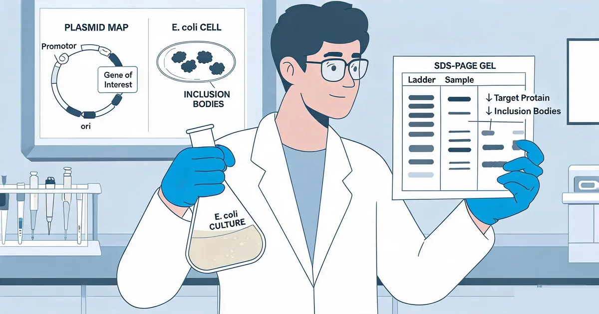
E. coli recombinant protein expression is the use of Escherichia coli bacteria as a host organism to manufacture a protein encoded by a foreign gene cloned into a plasmid vector. The gene is placed under control of an inducible promoter — most commonly the T7 promoter in pET-series vectors — so that expression can be switched on at a chosen point in the growth curve, typically with the inducer IPTG (isopropyl β-D-1-thiogalactopyranoside).
Compared to mammalian systems like CHO or HEK293, E. coli offers:
- Speed: Transformation to purified protein in 3–7 days versus 3–6 weeks for stable mammalian cell lines
- Cost: Simple, inexpensive growth media and no specialized cell culture infrastructure
- Yield: Gram-per-liter titers are achievable for well-behaved cytoplasmic proteins
The trade-off is that E. coli lacks the endoplasmic reticulum, chaperone diversity, and enzymatic machinery needed for glycosylation, complex disulfide bond formation, and many post-translational modifications. It is the right platform for enzymes, non-glycosylated antigens, and simple domains — and a poor fit for full-length human glycoproteins, which is why platforms like recombinant antigen expression systems comparing E. coli, CHO, insect, and yeast matter before you commit to a host.
Critical Principle
Every mistake in this article compounds the others. A poor codon-optimized gene expressed in the wrong strain, induced too aggressively, will fail in a way that looks identical to a fundamentally intractable target — even though the underlying protein might express perfectly well with three small changes.
2. Mistake 1: Ignoring Codon Usage Bias and mRNA Structure
The single most common — and most avoidable — mistake is submitting a gene sequence for synthesis or subcloning without checking it against E. coli's codon usage table. Genes from humans, viruses, or plants are written in the codon preferences of their native organism, and several of those codons are rare in E. coli's tRNA pool.
2.1 Why Rare Codons Break Expression
- Ribosome stalling: Codons like AGG, AGA, AUA, CUA, and CCC are decoded by low-abundance tRNAs in E. coli. Consecutive rare codons near the start of a gene stall the ribosome and cause premature termination or frameshifting.
- Truncated product: A stalled ribosome frequently produces a truncated protein that is invisible on a standard Coomassie-stained gel unless you specifically look for lower-molecular-weight bands.
- mRNA secondary structure: A strong hairpin loop within the first 30–40 nucleotides of the coding sequence can block ribosome binding entirely, independent of codon usage.
2.2 The Fix
- Run the gene sequence through a codon usage analysis tool before ordering synthesis
- Codon-optimize for E. coli, but avoid over-optimizing to a single high-frequency codon at every position — this can itself destabilize mRNA structure
- Check the first 15–20 codons specifically; rare codons here are more damaging than rare codons mid-sequence
- Consider a tRNA-supplemented strain (e.g., Rosetta, CodonPlus) as a fallback if resynthesis isn't feasible
"If a construct expresses nothing at all — not even a truncated band — check codon usage and mRNA folding before you touch the growth conditions."
3. Mistake 2: Wrong Strain, Vector, and Tag Combination
Teams often default to BL21(DE3) with an N-terminal His6 tag for every target, regardless of what the protein actually needs. That default works for a large fraction of constructs — but when it fails, changing the strain, tag, or vector backbone is usually a faster fix than redesigning the protein.
3.1 Strain Selection Matters
| Strain | Key Feature | Best For |
|---|---|---|
| BL21(DE3) | Chromosomal T7 RNA polymerase; protease-deficient (lon, ompT) | Default host for well-behaved, non-toxic targets |
| Rosetta(DE3) | Supplies tRNAs for 7 rare E. coli codons | Genes with significant rare-codon content |
| Origami / SHuffle | Oxidizing cytoplasm promotes disulfide bond formation | Proteins requiring intramolecular disulfide bonds |
| C41(DE3) / C43(DE3) | Tolerate toxic and membrane protein overexpression | Membrane proteins, toxic targets |
| Lemo21(DE3) | Tunable T7 RNA polymerase activity via L-rhamnose titration | Aggregation-prone targets needing slow expression |
3.2 Solubility Tags vs. Purification Tags
His6 and FLAG tags are excellent for affinity purification but do nothing to help a protein fold. When a target is prone to aggregation, fusing a solubility tag — MBP, SUMO, thioredoxin, or NusA — engages a well-folding partner protein that pulls the target through the folding pathway more successfully. SUMO fusions carry the added benefit of a protease (SUMO protease) that leaves a native N-terminus after cleavage, avoiding the extra residues left by many other tag-removal methods.
Common Mistake
Choosing a vector and tag combination based on what's already sitting on the bench, rather than what the target protein needs. A five-minute literature check on how homologous proteins have been expressed elsewhere often reveals the strain/tag combination that will save weeks of troubleshooting.
4. Mistake 3: Uncontrolled Induction Conditions
Standard protocols default to 1 mM IPTG at 37°C for 3–4 hours. For many targets, this is precisely the wrong recipe — it induces transcription faster than the cell's chaperone machinery can keep pace, driving the protein straight into inclusion bodies.
4.1 IPTG Concentration
- 1 mM (standard): Maximizes transcription rate; appropriate only for robust, fast-folding proteins
- 0.1–0.5 mM: A reasonable starting point for most novel targets
- 0.01–0.05 mM: Appropriate for aggregation-prone or multi-domain proteins where slow translation improves folding fidelity
4.2 Temperature
- 37°C: Fast growth, fast expression, highest aggregation risk
- 25–30°C: A common compromise — slower expression, meaningfully better solubility for many targets
- 15–18°C, overnight: Slowest expression kinetics; often the only condition that yields soluble protein for difficult targets, at the cost of a longer induction window (16–20 hours)
4.3 Induction Timing
Inducing too early (low OD600) wastes biomass; inducing too late (post-stationary phase) exposes the target to a nutrient-limited, protease-rich environment. Induce at OD600 0.6–0.8 for most constructs, and confirm by sampling pre- and post-induction time points on SDS-PAGE rather than assuming a single endpoint is representative.
Pro Tip
Run a 2×2×2 matrix — two IPTG concentrations, two temperatures, two harvest time points — in small-scale culture before committing to a fermentation run. It costs a day of bench time and routinely doubles or triples soluble yield.
5. Mistake 4: Underestimating Protein Toxicity
Some target proteins are directly toxic to E. coli — they interfere with membrane integrity, DNA replication, translation, or cell division. Symptoms of an unrecognized toxic construct include stalled or crashing cultures immediately after induction, rapid loss of plasmid (colonies losing antibiotic resistance), and an absence of the target band even when the gene sequence and codon usage are confirmed correct.
5.1 Recognizing Toxicity
- OD600 plateaus or drops shortly after induction, rather than continuing to climb
- Colonies appear only as satellite/small colonies on selective plates after transformation, suggesting leaky basal expression is already harming the host before induction
- Plasmid loss detected by re-streaking on selective media after a few generations without antibiotic pressure
5.2 Managing a Toxic Target
- Switch to a tightly repressed vector (pET with lacIq, or a T7 lysozyme-expressing strain such as BL21(DE3)pLysS) to suppress basal expression before induction
- Use C41(DE3)/C43(DE3), strains selected specifically for tolerance of toxic and membrane-disruptive proteins
- Lower induction temperature and IPTG concentration together to slow the rate of toxic protein accumulation
- Consider a fusion tag that sequesters the toxic domain, or express only a non-toxic truncated construct if the full-length protein is not required
6. Mistake 5: Skipping Small-Scale Optimization Before Scale-Up
Under project deadline pressure, it's tempting to jump straight from cloning confirmation to a large fermentation run. This is one of the costliest mistakes in E. coli recombinant protein expression: a failed 10-liter fermentation wastes days of instrument time, media, and labor that a 5 mL small-scale screen would have flagged in an afternoon.
- Screen before you scale: Test multiple strain/vector/induction combinations in 5–50 mL cultures before moving to shake-flask or fermenter scale.
- Confirm solubility, not just expression: A strong total-protein band on SDS-PAGE says nothing about whether the protein is soluble. Always compare soluble supernatant versus insoluble pellet fractions after lysis.
- Verify identity early: A band at the expected molecular weight is not confirmation — Western blot against the affinity tag or, for critical projects, intact mass spectrometry, should confirm identity before scale-up resources are committed.
IVD Application Note
For diagnostic-grade antigen production, document every small-scale optimization decision — strain, IPTG concentration, temperature, harvest time — as part of the process development record. Regulators and downstream QC teams will expect a rationale, and having comparative SDS-PAGE data on hand turns a documentation burden into a five-minute conversation.
7. Mistake 6: Treating Inclusion Bodies as Failure Instead of Data
Inclusion bodies — dense, insoluble aggregates of misfolded protein that accumulate in the E. coli cytoplasm — are often treated as a dead end. In reality, inclusion body formation is diagnostic information, and for some protein classes it's a viable production route rather than a failure mode.
7.1 What Inclusion Bodies Tell You
- The gene expresses well — inclusion bodies only form when transcription and translation are both functioning; the problem is downstream of expression, in folding kinetics.
- The mismatch is rate, not sequence — in most cases, slowing induction (lower IPTG, lower temperature) is sufficient to shift a meaningful fraction of the protein into the soluble phase.
7.2 Two Valid Paths Forward
- Push toward solubility: Lower IPTG and temperature, add a solubility tag (MBP/SUMO/thioredoxin), or co-express chaperone plasmids (e.g., GroEL/GroES, DnaK/DnaJ/GrpE).
- Embrace the inclusion bodies: For proteins with few or no disulfide bonds and simple secondary structure, denaturation with 6–8 M urea or guanidine hydrochloride followed by controlled dilution or dialysis refolding can recover high-purity, correctly folded protein — this is the standard route for many recombinant hormones and cytokines produced at industrial scale.
"Inclusion bodies aren't proof a protein can't be made in E. coli — they're proof it can be expressed. The only question left is whether refolding or a solubility-tag redesign gets you to native structure faster."
For antigens intended for immunoassay development, refolded material must be verified against native conformation-dependent antibody binding before it is accepted into a production workflow — a step easily skipped under schedule pressure but essential for reagents feeding sandwich ELISA or CLIA assay development.
8. Frequently Asked Questions — E. coli Recombinant Protein Expression Mistakes
What is E. coli recombinant protein expression?
E. coli recombinant protein expression is the use of Escherichia coli bacteria as a host to produce a protein encoded by a foreign gene inserted into a plasmid vector. It is the fastest and lowest-cost expression platform for non-glycosylated proteins, antigens, and enzymes, but it lacks the post-translational modification machinery of mammalian systems.
How long does it take to troubleshoot a failed E. coli expression construct?
A focused small-scale optimization screen — testing 2–3 strains, 2 temperatures, and 2 IPTG concentrations in parallel — typically takes 3–5 working days from transformation to SDS-PAGE readout. Diagnosing and redesigning around a codon bias or fusion tag problem can add another 1–2 weeks for gene resynthesis and subcloning.
Can I express a human membrane protein in E. coli?
It is possible but high-risk. Human membrane proteins depend on the Sec/YidC translocon, lipid composition, and often glycosylation for correct folding, none of which E. coli replicates natively. Specialized strains (C41/C43(DE3)) and slow, low-temperature induction improve success rates, but a mammalian system such as HEK293 is usually the more reliable choice for these targets.
What is the difference between a solubility tag and a purification tag?
A purification tag (His6, FLAG) exists solely to enable affinity capture and contributes little to folding. A solubility tag (MBP, SUMO, thioredoxin) is a larger, well-folding partner protein fused to the target that actively improves folding kinetics and cytoplasmic solubility. Many constructs use both: a solubility tag for expression, cleaved after purification, with a short affinity tag retained or removed as needed.
How do you evaluate whether a target protein is a good fit for E. coli expression?
Check three things before committing: whether the protein requires disulfide bonds or glycosylation for activity, whether it is toxic to bacterial membranes or DNA replication, and whether homologs from related organisms have been successfully expressed in E. coli in the literature. Small-scale expression trials across 2–3 constructs remain the fastest way to get a definitive answer.
Does Sekbio offer E. coli recombinant protein expression services?
Sekbio's core recombinant expression platform is built around CHO and HEK293 mammalian systems for antibodies, antigens, and fusion proteins that require native folding and glycosylation. For targets that are natively cytoplasmic, non-glycosylated, or better suited to bacterial hosts, our team evaluates the construct against all available expression systems and recommends the platform that will deliver the most reliable diagnostic-grade material. Visit our Recombinant Protein Expression Services page to discuss your target.
9. Summary
Most E. coli recombinant protein expression failures trace back to a small set of recurring mistakes:
- Codon usage bias: Check rare codons and mRNA secondary structure before synthesis, especially in the first 20 codons.
- Strain, vector, and tag mismatch: Match the host strain and fusion tag to what the specific target needs, not to what's already on the bench.
- Uncontrolled induction: Titrate IPTG concentration and temperature systematically rather than defaulting to 1 mM IPTG at 37°C.
- Unrecognized toxicity: Watch for culture crashes and plasmid loss as early warning signs, and switch to tightly repressed vectors or tolerant strains.
- Skipped small-scale screening: Always validate solubility and identity at 5–50 mL scale before committing to fermentation.
- Inclusion bodies: Treat them as diagnostic data, not dead ends — refolding or solubility-tag redesign are both valid recovery paths.
At Sekbio, our recombinant protein expression team evaluates every target against CHO, HEK293, and bacterial expression options before recommending a production route, so IVD developers get diagnostic-grade antigens and antibodies without repeating these avoidable mistakes. If you're evaluating expression systems for a new target, our Recombinant Protein Expression Services team can help you scope the right approach.