Anti-Müllerian hormone (AMH) has become the single most requested fertility hormone test worldwide — a one-time blood draw that estimates how many eggs a woman has left. But AMH is also one of the most misread numbers in reproductive medicine: patients frequently confuse "low AMH" with "infertile," and "high AMH" with "highly fertile." Neither is accurate. This guide covers what AMH actually measures, normal AMH levels by age, what low and high AMH mean clinically, how the AMH blood test works, and the real relationship between AMH and IVF success.
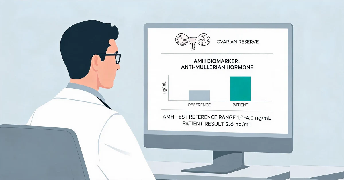
What Is AMH (Anti-Müllerian Hormone)?
AMH (anti-Müllerian hormone), also called Müllerian-inhibiting substance, is a glycoprotein hormone produced by the granulosa cells that surround small, growing (preantral and small antral) follicles in the ovary. Because the number of these early-stage follicles is proportional to a woman's total remaining egg supply, serum AMH concentration is used clinically as a marker of ovarian reserve — an estimate of egg quantity, not egg quality.
AMH has two properties that make it clinically convenient compared with other fertility hormones: it shows minimal fluctuation across the menstrual cycle, so it can be drawn on any day, and it is largely independent of the hypothalamic-pituitary-ovarian feedback loop that drives cycle-dependent swings in FSH, LH, and estradiol. AMH rises from birth, peaks around the mid-to-late 20s, then declines progressively through the 30s and 40s until it becomes undetectable roughly 5 years before the final menstrual period — making the AMH trajectory a biological clock for the ovarian follicle pool. (Source: Broer et al., Hum Reprod Update 2014)
Units note: AMH is most commonly reported in ng/mL in the United States and in pmol/L in Europe and parts of Asia. To convert, multiply ng/mL by 7.14 to get pmol/L (e.g., 2.0 ng/mL ≈ 14.3 pmol/L). Always confirm which unit and which assay generation your laboratory uses before comparing results across visits or clinics.
Normal AMH Levels by Age
AMH levels by age follow a well-documented, gradually declining curve. The table below reflects commonly used clinical reference ranges, consistent with large age-stratified cohort data. (Source: Seifer et al., Fertil Steril 2011 — a cohort of 17,120 women presenting to U.S. fertility centers)
| Age Group | Median AMH | Typical Range | Ovarian Reserve Interpretation |
|---|---|---|---|
| 20-24 years | ~3.8 ng/mL | 3.0-5.0 ng/mL | High / expected for age |
| 25-29 years | ~3.2 ng/mL | 2.5-4.0 ng/mL | Normal-high |
| 30-34 years | ~2.4 ng/mL | 1.8-3.0 ng/mL | Normal |
| 35-39 years | ~1.5 ng/mL | 1.0-2.0 ng/mL | Declining, expected for age |
| 40+ years | ~0.6 ng/mL | 0.3-1.0 ng/mL | Low, consistent with reduced reserve |
The average yearly decline in median AMH is approximately 0.2 ng/mL/year through age 35, slowing to roughly 0.1 ng/mL/year thereafter. Individual variation is substantial: two women of the same age can have AMH values that differ several-fold and both still fall within an expected range, which is why AMH is interpreted as a population-level trend rather than a precise individual prediction of remaining reproductive years.
Low AMH Levels — Causes & Fertility Impact
Low AMH — generally defined as below 1.0-1.1 ng/mL, with severely low AMH below 0.5 ng/mL — indicates diminished ovarian reserve (DOR): fewer remaining follicles than expected for age.
Common Causes of Low AMH
- Natural aging: The most common cause — AMH declines predictably as the follicle pool is depleted with each menstrual cycle
- Diminished ovarian reserve (DOR): Age-inappropriate decline, sometimes detected in women in their 20s or 30s
- Ovarian surgery: Cystectomy (especially for endometriomas) can remove healthy ovarian tissue alongside the cyst
- Chemotherapy or pelvic radiation: Gonadotoxic treatments can accelerate follicle loss
- Endometriosis: Associated with lower AMH, independent of surgical history
- Smoking: Associated with earlier ovarian aging and modestly lower AMH
- Primary ovarian insufficiency (POI): Very low or undetectable AMH before age 40
The clinical impact of low AMH is primarily on egg quantity retrievable during IVF stimulation, not on the ability to conceive naturally. A woman with low AMH who is still ovulating regularly and has open fallopian tubes can conceive spontaneously — low AMH lowers the odds and shortens the practical window, but it is not equivalent to infertility. It does, however, predict a lower egg yield and higher cycle cancellation risk if ovarian stimulation is pursued, which is why low-AMH patients are often counseled toward earlier evaluation and, if desired, earlier fertility treatment.
High AMH Levels — PCOS & Polycystic Ovaries
High AMH — typically above 3.5-4.0 ng/mL, or above the age-specific 95th percentile — usually reflects a higher-than-average number of small antral follicles. Elevated AMH is one of the most consistent hormonal findings in polycystic ovary syndrome (PCOS), where follicles accumulate at a small size but fail to mature and ovulate normally.
- PCOS association: AMH levels in women with PCOS are often 2-3× higher than age-matched controls, and some diagnostic algorithms now use AMH as a supportive criterion alongside irregular cycles and hyperandrogenism
- OHSS risk marker: High AMH going into IVF stimulation predicts a strong response and raises the risk of ovarian hyperstimulation syndrome (OHSS) — clinics typically respond with lower gonadotropin doses and antagonist protocols
- Not a fertility guarantee: High AMH indicates a large follicle pool, but PCOS-related anovulation can still make natural conception difficult without ovulation induction
Clinical note: AMH alone does not diagnose PCOS. Diagnosis requires the Rotterdam criteria — irregular ovulation, clinical or biochemical hyperandrogenism, and polycystic ovarian morphology on ultrasound (at least two of three). Elevated AMH is a strong supporting finding but should not replace a full clinical work-up.
AMH Test — How It Works & What to Expect
The AMH blood test (anti-Müllerian hormone test) is a simple serum draw with several practical advantages over other ovarian reserve markers:
- No cycle-day requirement: Unlike day-3 FSH, AMH can be drawn on any day of the menstrual cycle
- No fasting required
- Minimal intra-cycle variability: AMH shows far less fluctuation than FSH or estradiol across a single cycle
- Turnaround time: Results are typically available in 1-3 business days, depending on the assay platform
- Assay platforms: ELISA, chemiluminescent immunoassay (CLIA/ECLIA), and fluorescence immunoassay (FIA) are all used to quantify AMH, generally with a working range of approximately 0.01-20+ ng/mL
One important technical caveat for both clinicians and IVD developers: early-generation AMH ELISA kits were affected by complement-related sample interference, which could cause inconsistent results between assay generations and required revised sample handling protocols. Modern automated AMH immunoassays have largely resolved this issue, but it underscores why AMH results should always be compared using the same assay platform over time whenever possible, and why antibody pair specificity and sample pretreatment validation are critical in AMH IVD assay design. IVD developers can read our AMH antibody pair selection guide for the full epitope pairing and cross-reactivity screening process.
Fertility clinics increasingly pair central-lab AMH testing with point-of-care screening. See our AMH rapid test for fertility clinics overview for POCT-format AMH testing options.
AMH Levels & IVF Success Rates
AMH is the strongest single hormonal predictor of ovarian response to stimulation — how many eggs will likely be retrieved in an IVF cycle — and is used routinely to individualize gonadotropin starting doses. Higher AMH generally predicts a higher egg yield; lower AMH predicts a lower yield and a greater chance of cycle cancellation due to poor response.
What AMH Does and Does Not Predict in IVF
| Outcome | AMH Predictive Value |
|---|---|
| Number of eggs retrieved | Strong predictor |
| Gonadotropin dose needed | Strong predictor — used for protocol individualization |
| Risk of poor response / cancellation | Strong predictor when very low |
| Risk of OHSS | Strong predictor when very high |
| Egg / embryo quality | Weak — age is the dominant factor |
| Live birth rate per cycle | Weak-moderate — age remains the primary driver |
| Chance of natural conception | Weak — ovulation and tubal status matter more |
This distinction — AMH predicts quantity, not quality — is the single most important concept for patients to understand. A 38-year-old with high AMH will likely yield many eggs during stimulation, but her live birth rate per embryo remains governed by age-related aneuploidy risk. Conversely, a 30-year-old with low AMH may yield fewer eggs but often retains age-typical egg quality, meaning cumulative success across multiple stimulation cycles or accumulated embryos can still be favorable. AMH-based individualized dosing has been shown to improve cycle safety and reduce both under- and over-response compared with fixed-dose protocols. (Source: La Marca et al., Hum Reprod Update 2010)
AMH Antibody Pair for Fertility IVD Manufacturing
Sekbio supplies anti-AMH monoclonal antibody pairs for FIA and CLIA fertility assay development — ISO 13485 certified, validated across the full clinical AMH range for ovarian reserve and PCOS testing platforms.
Frequently Asked Questions About AMH
What is AMH (anti-Müllerian hormone)?
AMH is a protein hormone produced by granulosa cells surrounding small, growing follicles in the ovary. Serum AMH correlates with the number of these early-stage follicles, so it is used clinically as a marker of ovarian reserve — the size of a woman's remaining egg supply. AMH shows minimal fluctuation across the menstrual cycle and can be tested on any day.
AMH peaks in the mid-to-late 20s, declines gradually through the 30s and 40s, and becomes undetectable around menopause, roughly 5 years before the final menstrual period.
What is a normal AMH level by age?
Normal AMH declines steadily with age: approximately 3.0-5.0 ng/mL at 20-24 years, 2.5-4.0 ng/mL at 25-29, 1.8-3.0 ng/mL at 30-34, 1.0-2.0 ng/mL at 35-39, and 0.3-1.0 ng/mL at 40 and older.
These ranges vary between individuals of the same age and between assay platforms, so results should always be interpreted against the reporting laboratory's own reference range.
What is considered a low AMH level?
Most fertility clinics consider AMH below 1.0-1.1 ng/mL indicative of diminished ovarian reserve, with levels below 0.5 ng/mL considered severely low. Low AMH signals fewer remaining follicles and predicts a lower egg yield during IVF stimulation.
Low AMH reflects egg quantity, not egg quality or the ability to conceive naturally — many women with low AMH still conceive spontaneously if they are ovulating regularly.
What does high AMH mean, and is it linked to PCOS?
High AMH (typically above 3.5-4.0 ng/mL) usually reflects a higher-than-average number of small antral follicles and is one of the most consistent hormonal findings in polycystic ovary syndrome (PCOS).
Very high AMH is not itself dangerous, but during IVF stimulation it raises the risk of ovarian hyperstimulation syndrome (OHSS), which clinics manage with lower gonadotropin doses and antagonist protocols.
How is the AMH test performed?
The AMH test is a single blood draw that can be taken on any day of the menstrual cycle, with no fasting required. Samples are processed on ELISA or automated immunoassay platforms (CLIA, ECLIA, or FIA), reporting a quantitative concentration in ng/mL or pmol/L, usually within 1-3 business days.
Does AMH predict IVF success?
AMH is the best single hormonal predictor of how many eggs will be retrieved during IVF stimulation and is used to individualize gonadotropin dosing. It is a quantity marker, not a quality marker — it does not directly predict embryo quality or live birth rate, which are more strongly influenced by age.
Can AMH levels increase or be improved?
AMH reflects a fixed follicle pool established before birth, and no supplement or lifestyle change has been proven to increase the underlying follicle count once it has declined. Apparent increases are usually explained by assay variability, cycle timing, or resolution of temporary suppression from hormonal contraceptives.